 Movie
Movie Controller

[English] 日本語
 Yorodumi
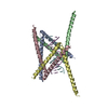
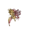
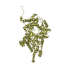
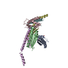
- PDB-5yti: Crystal structure of flagellar hook associated protein-3 (HAP-3: ... -
+ Open data

- Basic information
| Entry | Database: PDB / ID: 5yti | ||||||
|---|---|---|---|---|---|---|---|
| Title | Crystal structure of flagellar hook associated protein-3 (HAP-3: Q5ZW61_LEGPH) from Legionella pneumophila | ||||||
 Components Components | Flagellar hook associated protein type 3 FlgL | ||||||
 Keywords Keywords |  STRUCTURAL PROTEIN / Flagellar hook associated protein3 / HAP-3 / Legionella pneumophila STRUCTURAL PROTEIN / Flagellar hook associated protein3 / HAP-3 / Legionella pneumophila | ||||||
| Function / homology |  Function and homology information Function and homology informationbacterial-type flagellum hook / bacterial-type flagellum-dependent cell motility / structural molecule activity / extracellular region Similarity search - Function | ||||||
| Biological species |  Legionella pneumophila subsp. pneumophila (bacteria) Legionella pneumophila subsp. pneumophila (bacteria) | ||||||
| Method | X-RAY DIFFRACTION / SYNCHROTRON / AB INITIO PHASING / SAD / Resolution: 2.75 Å | ||||||
 Authors Authors | Lankipalli, S. / Hegde, R.P. / Dey, D. / Almo, S.C. / Ramagopal, U.A. | ||||||
 Citation Citation | Journal: To be published Title: Crystal structure of hook associated protein-3 (HAP-3: Q5ZW61_LEGPH) from Legionella pneumophila Authors: Lankipalli, S. / Ramagopal, U.A. | ||||||
| History |
|
- Structure visualization
| Structure viewer | Molecule: MolmilJmol/JSmol |
|---|
- Downloads & links
-Download
| PDBx/mmCIF format | 5yti.cif.gz | 135.9 KB | Display | PDBx/mmCIF format |
|---|---|---|---|---|
| PDB format | pdb5yti.ent.gz | 113.2 KB | Display | PDB format |
| PDBx/mmJSON format | 5yti.json.gz | Tree view | PDBx/mmJSON format | |
| Others |  Other downloads Other downloads |
-Validation report
| Arichive directory | https://data.pdbj.org/pub/pdb/validation_reports/yt/5ytiftp://data.pdbj.org/pub/pdb/validation_reports/yt/5yti | HTTPS FTP |
|---|
-Related structure data
| Similar structure data |
|---|


-Links
 PDBj
PDBj- Assembly
| Deposited unit | 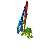
| ||||||||
|---|---|---|---|---|---|---|---|---|---|
| 1 | 
| ||||||||
| Unit cell |
| ||||||||
| Details | AUTHOR DETERMINED BIOLOGICAL UNIT: UNKNOWN |
-Components
| #1: Protein | Mass: 44940.734 Da / Num. of mol.: 1 Source method: isolated from a genetically manipulated source Source: (gene. exp.) Legionella pneumophila subsp. pneumophila (strain Philadelphia 1 / ATCC 33152 / DSM 7513) (bacteria)Strain: Philadelphia 1 / ATCC 33152 / DSM 7513 / Gene: flgL, lpg1226 / Plasmid: BC-PSGX4(BC) / Production host: Escherichia coli BL21(DE3) (bacteria) / Strain (production host): BL21(DE3) / References: UniProt: Q5ZW61 |
|---|---|
| #2: Chemical | ChemComp-CD /   Mass: 112.411 Da / Num. of mol.: 1 / Source method: obtained synthetically / Formula: Cd Mass: 112.411 Da / Num. of mol.: 1 / Source method: obtained synthetically / Formula: Cd |
| #3: Water | ChemComp-HOH / Water Mass: 18.015 Da / Num. of mol.: 2 / Source method: isolated from a natural source / Formula: H2O Mass: 18.015 Da / Num. of mol.: 2 / Source method: isolated from a natural source / Formula: H2O |
-Experimental details
-Experiment
| Experiment | Method: X-RAY DIFFRACTION / Number of used crystals: 1 |
|---|
- Sample preparation
| Crystal | Density Matthews: 2.91 Å3/Da / Density % sol: 57.79 % / Mosaicity: 0.294 ° |
|---|---|
| Crystal grow | Temperature: 298 K / Method: vapor diffusion, sitting drop / pH: 4.6 Details: 30% PEG 400, 0.1M Cadmium Chloride hydrate, 0.1M Sodium Acetate Trihydrate, pH 4.6 |
-Data collection
| Diffraction | Mean temperature: 100 K | |||||||||||||||||||||||||||||||||||||||||||||||||||||||||||||||||||||||||||||||||||||||||||||||||||||||||||||||||||||||||||||||||||||||||||||||||||||||||||||||||||||||||||||||||||||||||||||
|---|---|---|---|---|---|---|---|---|---|---|---|---|---|---|---|---|---|---|---|---|---|---|---|---|---|---|---|---|---|---|---|---|---|---|---|---|---|---|---|---|---|---|---|---|---|---|---|---|---|---|---|---|---|---|---|---|---|---|---|---|---|---|---|---|---|---|---|---|---|---|---|---|---|---|---|---|---|---|---|---|---|---|---|---|---|---|---|---|---|---|---|---|---|---|---|---|---|---|---|---|---|---|---|---|---|---|---|---|---|---|---|---|---|---|---|---|---|---|---|---|---|---|---|---|---|---|---|---|---|---|---|---|---|---|---|---|---|---|---|---|---|---|---|---|---|---|---|---|---|---|---|---|---|---|---|---|---|---|---|---|---|---|---|---|---|---|---|---|---|---|---|---|---|---|---|---|---|---|---|---|---|---|---|---|---|---|---|---|---|---|
| Diffraction source | Source: SYNCHROTRON / Site: NSLS  / Beamline: X29A / Wavelength: 0.987 Å / Beamline: X29A / Wavelength: 0.987 Å | |||||||||||||||||||||||||||||||||||||||||||||||||||||||||||||||||||||||||||||||||||||||||||||||||||||||||||||||||||||||||||||||||||||||||||||||||||||||||||||||||||||||||||||||||||||||||||||
| Detector | Type: ADSC QUANTUM 315 / Detector: CCD / Date: Jun 22, 2013 | |||||||||||||||||||||||||||||||||||||||||||||||||||||||||||||||||||||||||||||||||||||||||||||||||||||||||||||||||||||||||||||||||||||||||||||||||||||||||||||||||||||||||||||||||||||||||||||
| Radiation | Protocol: SINGLE WAVELENGTH / Monochromatic (M) / Laue (L): M / Scattering type: x-ray | |||||||||||||||||||||||||||||||||||||||||||||||||||||||||||||||||||||||||||||||||||||||||||||||||||||||||||||||||||||||||||||||||||||||||||||||||||||||||||||||||||||||||||||||||||||||||||||
| Radiation wavelength | Wavelength: 0.987 Å / Relative weight: 1 | |||||||||||||||||||||||||||||||||||||||||||||||||||||||||||||||||||||||||||||||||||||||||||||||||||||||||||||||||||||||||||||||||||||||||||||||||||||||||||||||||||||||||||||||||||||||||||||
| Reflection | Resolution: 2.75→76.15 Å / Num. obs: 14351 / % possible obs: 100 % / Redundancy: 8.7 % / Rmerge(I) obs: 0.089 / Rpim(I) all: 0.032 / Rrim(I) all: 0.094 / Χ2: 0.884 / Net I/σ(I): 7.5 | |||||||||||||||||||||||||||||||||||||||||||||||||||||||||||||||||||||||||||||||||||||||||||||||||||||||||||||||||||||||||||||||||||||||||||||||||||||||||||||||||||||||||||||||||||||||||||||
| Reflection shell | Diffraction-ID: 1
|
-Phasing
| Phasing | Method: SAD |
|---|
- Processing
| Software |
| |||||||||||||||||||||||||||||||||||||||||||||||||||||||||||||||||||||||||||
|---|---|---|---|---|---|---|---|---|---|---|---|---|---|---|---|---|---|---|---|---|---|---|---|---|---|---|---|---|---|---|---|---|---|---|---|---|---|---|---|---|---|---|---|---|---|---|---|---|---|---|---|---|---|---|---|---|---|---|---|---|---|---|---|---|---|---|---|---|---|---|---|---|---|---|---|---|
| Refinement | Method to determine structure: AB INITIO PHASING / Resolution: 2.75→76.15 Å / Cor.coef. Fo:Fc: 0.918 / Cor.coef. Fo:Fc free: 0.888 / SU B: 36.065 / SU ML: 0.3 / SU R Cruickshank DPI: 0.5507 / Cross valid method: THROUGHOUT / σ(F): 0 / ESU R: 0.551 / ESU R Free: 0.347 Details: HYDROGENS HAVE BEEN ADDED IN THE RIDING POSITIONS U VALUES : WITH TLS ADDED
| |||||||||||||||||||||||||||||||||||||||||||||||||||||||||||||||||||||||||||
| Solvent computation | Ion probe radii: 0.8 Å / Shrinkage radii: 0.8 Å / VDW probe radii: 1.2 Å | |||||||||||||||||||||||||||||||||||||||||||||||||||||||||||||||||||||||||||
| Displacement parameters | Biso max: 153.61 Å2 / Biso mean: 91.848 Å2 / Biso min: 62.07 Å2
| |||||||||||||||||||||||||||||||||||||||||||||||||||||||||||||||||||||||||||
| Refinement step | Cycle: final / Resolution: 2.75→76.15 Å
| |||||||||||||||||||||||||||||||||||||||||||||||||||||||||||||||||||||||||||
| Refine LS restraints |
| |||||||||||||||||||||||||||||||||||||||||||||||||||||||||||||||||||||||||||
| LS refinement shell | Resolution: 2.75→2.821 Å / Rfactor Rfree error: 0 / Total num. of bins used: 20
| |||||||||||||||||||||||||||||||||||||||||||||||||||||||||||||||||||||||||||
| Refinement TLS params. | Method: refined / Refine-ID: X-RAY DIFFRACTION
| |||||||||||||||||||||||||||||||||||||||||||||||||||||||||||||||||||||||||||
| Refinement TLS group |
|
