 Movie
Movie Controller

[English] 日本語
 Yorodumi
- PDB-5ysq: Sulfate-complex structure of a pyrophosphate-dependent kinase in ... -
+ Open data

- Basic information
| Entry | Database: PDB / ID: 5ysq | |||||||||
|---|---|---|---|---|---|---|---|---|---|---|
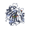
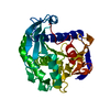
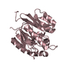
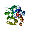
| Title | Sulfate-complex structure of a pyrophosphate-dependent kinase in the ribokinase family provides insight into the donor-binding mode | |||||||||
 Components Components | TM0415 | |||||||||
 Keywords Keywords |  TRANSFERASE TRANSFERASE | |||||||||
| Function / homology |  Function and homology information Function and homology information | |||||||||
| Biological species |   Thermotoga maritima (bacteria) Thermotoga maritima (bacteria) | |||||||||
| Method | X-RAY DIFFRACTION / SYNCHROTRON / MOLECULAR REPLACEMENT / Resolution: 1.47 Å | |||||||||
 Authors Authors | Nagata, R. / Fujihashi, M. / Miki, K. | |||||||||
| Funding support |  Japan, 2items Japan, 2items
| |||||||||
 Citation Citation | Journal: Nat Commun / Year: 2018 Title: Identification of a pyrophosphate-dependent kinase and its donor selectivity determinants. Authors: Nagata, R. / Fujihashi, M. / Sato, T. / Atomi, H. / Miki, K. | |||||||||
| History |
|
- Structure visualization
| Structure viewer | Molecule: MolmilJmol/JSmol |
|---|
- Downloads & links
-Download
| PDBx/mmCIF format | 5ysq.cif.gz | 237.6 KB | Display | PDBx/mmCIF format |
|---|---|---|---|---|
| PDB format | pdb5ysq.ent.gz | 191.8 KB | Display | PDB format |
| PDBx/mmJSON format | 5ysq.json.gz | Tree view | PDBx/mmJSON format | |
| Others |  Other downloads Other downloads |
-Validation report
| Arichive directory | https://data.pdbj.org/pub/pdb/validation_reports/ys/5ysqftp://data.pdbj.org/pub/pdb/validation_reports/ys/5ysq | HTTPS FTP |
|---|
-Related structure data
| Related structure data |  5yspC  1vk4S S: Starting model for refinement C: citing same article ( |
|---|---|
| Similar structure data |


-Links
 PDBj
PDBj- Assembly
| Deposited unit | 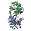
| ||||||||
|---|---|---|---|---|---|---|---|---|---|
| 1 | 
| ||||||||
| 2 | 
| ||||||||
| Unit cell |
|
-Components
| #1: Protein | Mass: 32104.656 Da / Num. of mol.: 2 Source method: isolated from a genetically manipulated source Source: (gene. exp.) Thermotoga maritima (strain ATCC 43589 / MSB8 / DSM 3109 / JCM 10099) (bacteria)Strain: ATCC 43589 / MSB8 / DSM 3109 / JCM 10099 / Gene: TM_0415 / Plasmid: pET-15b / Production host: Escherichia coli (E. coli) / Strain (production host): BL21(DE3)pLysS / References: UniProt: Q9WYP6#2: Chemical | Inositol  Mass: 180.156 Da / Num. of mol.: 2 / Source method: obtained synthetically / Formula: C6H12O6 / Comment: neurotransmitter, hormone*YM Mass: 180.156 Da / Num. of mol.: 2 / Source method: obtained synthetically / Formula: C6H12O6 / Comment: neurotransmitter, hormone*YM#3: Chemical | ChemComp-SO4 / Sulfate  Mass: 96.063 Da / Num. of mol.: 4 / Source method: obtained synthetically / Formula: SO4 / Feature type: SUBJECT OF INVESTIGATION Mass: 96.063 Da / Num. of mol.: 4 / Source method: obtained synthetically / Formula: SO4 / Feature type: SUBJECT OF INVESTIGATION#4: Water | ChemComp-HOH / | Water Mass: 18.015 Da / Num. of mol.: 258 / Source method: isolated from a natural source / Formula: H2O Mass: 18.015 Da / Num. of mol.: 258 / Source method: isolated from a natural source / Formula: H2O |
|---|
-Experimental details
-Experiment
| Experiment | Method: X-RAY DIFFRACTION / Number of used crystals: 1 |
|---|
- Sample preparation
| Crystal | Density Matthews: 1.94 Å3/Da / Density % sol: 36.53 % |
|---|---|
| Crystal grow | Temperature: 293 K / Method: vapor diffusion, sitting drop / pH: 4.6 / Details: PEG 4000, ammonium sulfate, sodium acetate |
-Data collection
| Diffraction | Mean temperature: 100 K |
|---|---|
| Diffraction source | Source: SYNCHROTRON / Site: SPring-8 / Beamline: BL41XU / Wavelength: 1 Å |
| Detector | Type: DECTRIS PILATUS3 6M / Detector: PIXEL / Date: Oct 8, 2016 |
| Radiation | Protocol: SINGLE WAVELENGTH / Monochromatic (M) / Laue (L): M / Scattering type: x-ray |
| Radiation wavelength | Wavelength: 1 Å / Relative weight: 1 |
| Reflection | Resolution: 1.47→50 Å / Num. obs: 79847 / % possible obs: 95.7 % / Redundancy: 3.4 % / Net I/σ(I): 27.8 |
| Reflection shell | Resolution: 1.47→1.5 Å |
- Processing
| Software |
| ||||||||||||||||||||||||||||||||||||||||||||||||||||||||||||||||||||||||||||||||||||||||||||||||||||||||||||||||||||||||||||||||||||||||||||||||||||||||||||||||||||||||||||||||||||||
|---|---|---|---|---|---|---|---|---|---|---|---|---|---|---|---|---|---|---|---|---|---|---|---|---|---|---|---|---|---|---|---|---|---|---|---|---|---|---|---|---|---|---|---|---|---|---|---|---|---|---|---|---|---|---|---|---|---|---|---|---|---|---|---|---|---|---|---|---|---|---|---|---|---|---|---|---|---|---|---|---|---|---|---|---|---|---|---|---|---|---|---|---|---|---|---|---|---|---|---|---|---|---|---|---|---|---|---|---|---|---|---|---|---|---|---|---|---|---|---|---|---|---|---|---|---|---|---|---|---|---|---|---|---|---|---|---|---|---|---|---|---|---|---|---|---|---|---|---|---|---|---|---|---|---|---|---|---|---|---|---|---|---|---|---|---|---|---|---|---|---|---|---|---|---|---|---|---|---|---|---|---|---|---|
| Refinement | Method to determine structure: MOLECULAR REPLACEMENT Starting model: 1VK4 Resolution: 1.47→50 Å / Cor.coef. Fo:Fc: 0.958 / Cor.coef. Fo:Fc free: 0.956 / SU B: 3.043 / SU ML: 0.056 / Cross valid method: THROUGHOUT / ESU R: 0.085 / ESU R Free: 0.078 / Details: HYDROGENS HAVE BEEN ADDED IN THE RIDING POSITIONS
| ||||||||||||||||||||||||||||||||||||||||||||||||||||||||||||||||||||||||||||||||||||||||||||||||||||||||||||||||||||||||||||||||||||||||||||||||||||||||||||||||||||||||||||||||||||||
| Solvent computation | Ion probe radii: 0.8 Å / Shrinkage radii: 0.8 Å / VDW probe radii: 1.2 Å | ||||||||||||||||||||||||||||||||||||||||||||||||||||||||||||||||||||||||||||||||||||||||||||||||||||||||||||||||||||||||||||||||||||||||||||||||||||||||||||||||||||||||||||||||||||||
| Displacement parameters | Biso mean: 17.497 Å2
| ||||||||||||||||||||||||||||||||||||||||||||||||||||||||||||||||||||||||||||||||||||||||||||||||||||||||||||||||||||||||||||||||||||||||||||||||||||||||||||||||||||||||||||||||||||||
| Refinement step | Cycle: 1 / Resolution: 1.47→50 Å
| ||||||||||||||||||||||||||||||||||||||||||||||||||||||||||||||||||||||||||||||||||||||||||||||||||||||||||||||||||||||||||||||||||||||||||||||||||||||||||||||||||||||||||||||||||||||
| Refine LS restraints |
|