 Movie
Movie Controller

[English] 日本語
 Yorodumi
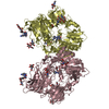
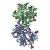
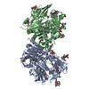
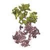
- PDB-5yp4: Crystal structure of dipeptidyl peptidase IV (DPP IV) with Lys-Pr... -
+ Open data

- Basic information
| Entry | Database: PDB / ID: 5yp4 | |||||||||||||||||||||||||||||||||
|---|---|---|---|---|---|---|---|---|---|---|---|---|---|---|---|---|---|---|---|---|---|---|---|---|---|---|---|---|---|---|---|---|---|---|
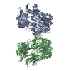
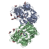
| Title | Crystal structure of dipeptidyl peptidase IV (DPP IV) with Lys-Pro from Pseudoxanthomonas mexicana WO24 | |||||||||||||||||||||||||||||||||
 Components Components | Dipeptidyl aminopeptidase 4 Dipeptidyl peptidase Dipeptidyl peptidase | |||||||||||||||||||||||||||||||||
 Keywords Keywords | HYDROLASE / DAP IV / Clan SC S9 / peptidase / DPP4 / DPP8 / DPP9 | |||||||||||||||||||||||||||||||||
| Function / homology |  Function and homology informationdipeptidyl-peptidase IV / dipeptidyl-peptidase activity / aminopeptidase activity / serine-type peptidase activity / proteolysis involved in protein catabolic process / periplasmic space / protein homodimerization activity / identical protein binding / cytoplasm Function and homology informationdipeptidyl-peptidase IV / dipeptidyl-peptidase activity / aminopeptidase activity / serine-type peptidase activity / proteolysis involved in protein catabolic process / periplasmic space / protein homodimerization activity / identical protein binding / cytoplasmSimilarity search - Function | |||||||||||||||||||||||||||||||||
| Biological species |  Pseudoxanthomonas mexicana (bacteria) Pseudoxanthomonas mexicana (bacteria) | |||||||||||||||||||||||||||||||||
| Method | X-RAY DIFFRACTION / SYNCHROTRON / MOLECULAR REPLACEMENT / Resolution: 1.9 Å | |||||||||||||||||||||||||||||||||
 Authors Authors | Roppongi, S. / Suzuki, Y. / Tateoka, C. / Fuimoto, M. / Morisawa, S. / Iizuka, I. / Nakamura, A. / Honma, N. / Shida, Y. / Ogasawara, W. ...Roppongi, S. / Suzuki, Y. / Tateoka, C. / Fuimoto, M. / Morisawa, S. / Iizuka, I. / Nakamura, A. / Honma, N. / Shida, Y. / Ogasawara, W. / Tanaka, N. / Sakamoto, Y. / Nonaka, T. | |||||||||||||||||||||||||||||||||
| Funding support |  Japan, 10items Japan, 10items
| |||||||||||||||||||||||||||||||||
 Citation Citation | Journal: Sci Rep / Year: 2018 Title: Crystal structures of a bacterial dipeptidyl peptidase IV reveal a novel substrate recognition mechanism distinct from that of mammalian orthologues. Authors: Roppongi, S. / Suzuki, Y. / Tateoka, C. / Fujimoto, M. / Morisawa, S. / Iizuka, I. / Nakamura, A. / Honma, N. / Shida, Y. / Ogasawara, W. / Tanaka, N. / Sakamoto, Y. / Nonaka, T. | |||||||||||||||||||||||||||||||||
| History |
|
- Structure visualization
| Structure viewer | Molecule: MolmilJmol/JSmol |
|---|
- Downloads & links
-Download
| PDBx/mmCIF format | 5yp4.cif.gz | 616.6 KB | Display | PDBx/mmCIF format |
|---|---|---|---|---|
| PDB format | pdb5yp4.ent.gz | 503.9 KB | Display | PDB format |
| PDBx/mmJSON format | 5yp4.json.gz | Tree view | PDBx/mmJSON format | |
| Others |  Other downloads Other downloads |
-Validation report
| Arichive directory | https://data.pdbj.org/pub/pdb/validation_reports/yp/5yp4ftp://data.pdbj.org/pub/pdb/validation_reports/yp/5yp4 | HTTPS FTP |
|---|
-Related structure data
| Related structure data | 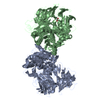 5yp1SC 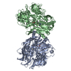 5yp2C  5yp3C S: Starting model for refinement C: citing same article ( |
|---|---|
| Similar structure data |


-Links
 PDBj
PDBj

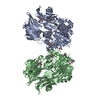
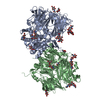
- Assembly
| Deposited unit | 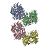
| ||||||||
|---|---|---|---|---|---|---|---|---|---|
| 1 | 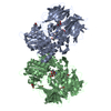
| ||||||||
| 2 | 
| ||||||||
| Unit cell |
|
-Components
| #1: Protein | Dipeptidyl peptidase / Dipeptidyl aminopeptidase IV / DAP IV Mass: 82375.500 Da / Num. of mol.: 4 Source method: isolated from a genetically manipulated source Source: (gene. exp.) Pseudoxanthomonas mexicana (bacteria) / Strain: WO24 / Gene: dap4 / Plasmid: pUC19 / Production host: Escherichia coli K-12 (bacteria) / Strain (production host): K-12 / References: UniProt: Q6F3I7, dipeptidyl-peptidase IV#2: Chemical | ChemComp-GOL / Glycerol  Mass: 92.094 Da / Num. of mol.: 21 / Source method: obtained synthetically / Formula: C3H8O3 Mass: 92.094 Da / Num. of mol.: 21 / Source method: obtained synthetically / Formula: C3H8O3#3: Chemical | ChemComp-LYS / Lysine  Type: L-peptide linking / Mass: 147.195 Da / Num. of mol.: 4 / Source method: obtained synthetically / Formula: C6H15N2O2 Type: L-peptide linking / Mass: 147.195 Da / Num. of mol.: 4 / Source method: obtained synthetically / Formula: C6H15N2O2#4: Chemical | ChemComp-PRO / Proline  Type: L-peptide linking / Mass: 115.130 Da / Num. of mol.: 4 / Source method: obtained synthetically / Formula: C5H9NO2 Type: L-peptide linking / Mass: 115.130 Da / Num. of mol.: 4 / Source method: obtained synthetically / Formula: C5H9NO2#5: Water | ChemComp-HOH / | Water Mass: 18.015 Da / Num. of mol.: 2877 / Source method: isolated from a natural source / Formula: H2O Mass: 18.015 Da / Num. of mol.: 2877 / Source method: isolated from a natural source / Formula: H2OSequence details | Residue M12I is mutagenesis according to sequence database UniportKB Q6F3I7 (DAP4_PSEMX). | |
|---|
-Experimental details
-Experiment
| Experiment | Method: X-RAY DIFFRACTION / Number of used crystals: 1 |
|---|
- Sample preparation
| Crystal | Density Matthews: 2.59 Å3/Da / Density % sol: 52.45 % |
|---|---|
| Crystal grow | Temperature: 293.15 K / Method: vapor diffusion, hanging drop / pH: 6.5 Details: 12% PEG 20000, 80mM MES pH6.5, 20% Glycerol, 4mM Lys-Pro |
-Data collection
| Diffraction | Mean temperature: 95 K |
|---|---|
| Diffraction source | Source: SYNCHROTRON / Site: Photon Factory / Beamline: BL-17A / Wavelength: 0.98 Å |
| Detector | Type: ADSC QUANTUM 270 / Detector: CCD / Date: Nov 20, 2013 |
| Radiation | Protocol: SINGLE WAVELENGTH / Monochromatic (M) / Laue (L): M / Scattering type: x-ray |
| Radiation wavelength | Wavelength: 0.98 Å / Relative weight: 1 |
| Reflection | Resolution: 1.9→50 Å / Num. obs: 245086 / % possible obs: 94.2 % / Redundancy: 2.2 % / Rmerge(I) obs: 0.082 / Rpim(I) all: 0.073 / Rrim(I) all: 0.11 / Net I/σ(I): 6.5 |
| Reflection shell | Resolution: 1.9→2 Å / Redundancy: 2.2 % / Rmerge(I) obs: 0.316 / Mean I/σ(I) obs: 2 / Num. unique obs: 27199 / Rpim(I) all: 0.282 / Rrim(I) all: 0.425 / % possible all: 71.5 |
- Processing
| Software |
| ||||||||||||||||||||||||||||||||||||||||||||||||||||||||||||||||||||||||||||||||||||||||||||||||||||||||||||||||||||||||||||||||||||||||||||||||||||||||||||||||||||||||||||||||||||||
|---|---|---|---|---|---|---|---|---|---|---|---|---|---|---|---|---|---|---|---|---|---|---|---|---|---|---|---|---|---|---|---|---|---|---|---|---|---|---|---|---|---|---|---|---|---|---|---|---|---|---|---|---|---|---|---|---|---|---|---|---|---|---|---|---|---|---|---|---|---|---|---|---|---|---|---|---|---|---|---|---|---|---|---|---|---|---|---|---|---|---|---|---|---|---|---|---|---|---|---|---|---|---|---|---|---|---|---|---|---|---|---|---|---|---|---|---|---|---|---|---|---|---|---|---|---|---|---|---|---|---|---|---|---|---|---|---|---|---|---|---|---|---|---|---|---|---|---|---|---|---|---|---|---|---|---|---|---|---|---|---|---|---|---|---|---|---|---|---|---|---|---|---|---|---|---|---|---|---|---|---|---|---|---|
| Refinement | Method to determine structure: MOLECULAR REPLACEMENT Starting model: 5YP1 Resolution: 1.9→40 Å / Cor.coef. Fo:Fc: 0.954 / Cor.coef. Fo:Fc free: 0.924 / SU B: 3.336 / SU ML: 0.098 / Cross valid method: THROUGHOUT / ESU R: 0.142 / ESU R Free: 0.139 / Stereochemistry target values: MAXIMUM LIKELIHOOD / Details: HYDROGENS HAVE BEEN ADDED IN THE RIDING POSITIONS
| ||||||||||||||||||||||||||||||||||||||||||||||||||||||||||||||||||||||||||||||||||||||||||||||||||||||||||||||||||||||||||||||||||||||||||||||||||||||||||||||||||||||||||||||||||||||
| Solvent computation | Ion probe radii: 0.8 Å / Shrinkage radii: 0.8 Å / VDW probe radii: 1.2 Å / Solvent model: MASK | ||||||||||||||||||||||||||||||||||||||||||||||||||||||||||||||||||||||||||||||||||||||||||||||||||||||||||||||||||||||||||||||||||||||||||||||||||||||||||||||||||||||||||||||||||||||
| Displacement parameters | Biso mean: 22.731 Å2
| ||||||||||||||||||||||||||||||||||||||||||||||||||||||||||||||||||||||||||||||||||||||||||||||||||||||||||||||||||||||||||||||||||||||||||||||||||||||||||||||||||||||||||||||||||||||
| Refinement step | Cycle: 1 / Resolution: 1.9→40 Å
| ||||||||||||||||||||||||||||||||||||||||||||||||||||||||||||||||||||||||||||||||||||||||||||||||||||||||||||||||||||||||||||||||||||||||||||||||||||||||||||||||||||||||||||||||||||||
| Refine LS restraints |
|