 Movie
Movie Controller


+ Open data

- Basic information
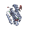
| Entry | Database: PDB / ID: 5ymx | ||||||
|---|---|---|---|---|---|---|---|
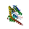
| Title | Myxococcus xanthus MglA in GDP bound conformation | ||||||
 Components Components | Mutual gliding-motility protein MglA | ||||||
 Keywords Keywords |  SIGNALING PROTEIN / HYDROLASE / Small Ras-like GTPase / Cytosolic / Myxococcus motility protein / spatial oscillation SIGNALING PROTEIN / HYDROLASE / Small Ras-like GTPase / Cytosolic / Myxococcus motility protein / spatial oscillation | ||||||
| Function / homology |  Function and homology informationregulation of protein localization / GTPase activity / GTP binding / cytoplasm Function and homology informationregulation of protein localization / GTPase activity / GTP binding / cytoplasmSimilarity search - Function | ||||||
| Biological species |  Myxococcus xanthus (bacteria) Myxococcus xanthus (bacteria) | ||||||
| Method | X-RAY DIFFRACTION / SYNCHROTRON / MOLECULAR REPLACEMENT / Resolution: 1.35 Å | ||||||
 Authors Authors | Baranwal, J. / Gayathri, P. | ||||||
| Funding support |  India, 1items India, 1items
| ||||||
 Citation Citation | Journal: Plos Biol. / Year: 2019 Title: Allosteric regulation of a prokaryotic small Ras-like GTPase contributes to cell polarity oscillations in bacterial motility. Authors: Baranwal, J. / Lhospice, S. / Kanade, M. / Chakraborty, S. / Gade, P.R. / Harne, S. / Herrou, J. / Mignot, T. / Gayathri, P. | ||||||
| History |
|
- Structure visualization
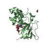
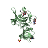
| Structure viewer | Molecule: MolmilJmol/JSmol |
|---|
- Downloads & links
-Download
| PDBx/mmCIF format | 5ymx.cif.gz | 199.1 KB | Display | PDBx/mmCIF format |
|---|---|---|---|---|
| PDB format | pdb5ymx.ent.gz | 158.4 KB | Display | PDB format |
| PDBx/mmJSON format | 5ymx.json.gz | Tree view | PDBx/mmJSON format | |
| Others |  Other downloads Other downloads |
-Validation report
| Arichive directory | https://data.pdbj.org/pub/pdb/validation_reports/ym/5ymxftp://data.pdbj.org/pub/pdb/validation_reports/ym/5ymx | HTTPS FTP |
|---|
-Related structure data
| Related structure data |  6izwC  3t12S S: Starting model for refinement C: citing same article ( |
|---|---|
| Similar structure data |


-Links
 PDBj
PDBj


- Assembly
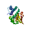
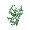
| Deposited unit | 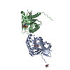
| ||||||||
|---|---|---|---|---|---|---|---|---|---|
| 1 | 
| ||||||||
| 2 | 
| ||||||||
| Unit cell |
|
-Components
-Protein , 1 types, 2 molecules AB
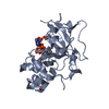
| #1: Protein | Mass: 23015.391 Da / Num. of mol.: 2 Source method: isolated from a genetically manipulated source Source: (gene. exp.) Myxococcus xanthus (strain DK 1622) (bacteria)Strain: DK 1622 / Gene: mglA, MXAN_1925 / Plasmid: pHis17 / Production host: Escherichia coli BL21(DE3) (bacteria) / Strain (production host): BL21(DE3) / References: UniProt: Q1DB04, small monomeric GTPase |
|---|
-Non-polymers , 5 types, 431 molecules 


| #2: Chemical | Guanosine diphosphate Type: RNA linking / Mass: 443.201 Da / Num. of mol.: 2 Type: RNA linking / Mass: 443.201 Da / Num. of mol.: 2Source method: isolated from a genetically manipulated source Formula: C10H15N5O11P2 / Comment: GDP, energy-carrying molecule*YM #3: Chemical | Imidazole Mass: 69.085 Da / Num. of mol.: 2 / Source method: obtained synthetically / Formula: C3H5N2 Mass: 69.085 Da / Num. of mol.: 2 / Source method: obtained synthetically / Formula: C3H5N2#4: Chemical | Ethylene glycol Mass: 62.068 Da / Num. of mol.: 2 / Source method: obtained synthetically / Formula: C2H6O2 Mass: 62.068 Da / Num. of mol.: 2 / Source method: obtained synthetically / Formula: C2H6O2#5: Chemical | ChemComp-MPD / ( | 2-Methyl-2,4-pentanediol Mass: 118.174 Da / Num. of mol.: 1 / Source method: obtained synthetically / Formula: C6H14O2 / Comment: precipitant*YM Mass: 118.174 Da / Num. of mol.: 1 / Source method: obtained synthetically / Formula: C6H14O2 / Comment: precipitant*YM#6: Water | ChemComp-HOH / | WaterMass: 18.015 Da / Num. of mol.: 424 / Source method: isolated from a natural source / Formula: H2O |
|---|
-Experimental details
-Experiment
| Experiment | Method: X-RAY DIFFRACTION / Number of used crystals: 1 |
|---|
- Sample preparation
| Crystal | Density Matthews: 2.35 Å3/Da / Density % sol: 47.63 % |
|---|---|
| Crystal grow | Temperature: 291 K / Method: vapor diffusion, sitting drop Details: 30% 2-Methyl 2,4-pentane diol (MPD), 10% PEG 4000, 100mM Imidazole, pH 8.0 PH range: 6.5 - 8.0 / Temp details: plates kept in an incubator at 291 K |
-Data collection
| Diffraction | Mean temperature: 100 K / Ambient temp details: nil |
|---|---|
| Diffraction source | Source: SYNCHROTRON / Site: ESRF  / Beamline: BM14 / Wavelength: 0.97625 Å / Beamline: BM14 / Wavelength: 0.97625 Å |
| Detector | Type: MARMOSAIC 225 mm CCD / Detector: CCD / Date: Jul 9, 2014 / Details: Bent collimating mirror and torroid |
| Radiation | Monochromator: Si(111) / Protocol: SINGLE WAVELENGTH / Monochromatic (M) / Laue (L): M / Scattering type: x-ray |
| Radiation wavelength | Wavelength: 0.97625 Å / Relative weight: 1 |
| Reflection | Resolution: 1.35→32.03 Å / Num. obs: 91269 / % possible obs: 99.7 % / Redundancy: 7.2 % / Biso Wilson estimate: 15.2 Å2 / CC1/2: 0.999 / Rmerge(I) obs: 0.055 / Rpim(I) all: 0.022 / Rrim(I) all: 0.06 / Rsym value: 0.06 / Net I/av σ(I): 8 / Net I/σ(I): 14.6 |
| Reflection shell | Resolution: 1.35→1.42 Å / Redundancy: 7.2 % / Rmerge(I) obs: 0.734 / Mean I/σ(I) obs: 2.6 / Num. unique obs: 13188 / CC1/2: 0.784 / Rpim(I) all: 0.294 / Rrim(I) all: 0.791 / Rsym value: 0.791 / % possible all: 100 |
- Processing
| Software |
| |||||||||||||||||||||||||||||||||||||||||||||||||||||||||||||||||||||||||||||||||||||||||||||||||||||||||||||||||||||||||||||||||||||||||||||||||||||||||||||||||||||||||||||||||||||||||||||||||||||||||||||||||||||||||
|---|---|---|---|---|---|---|---|---|---|---|---|---|---|---|---|---|---|---|---|---|---|---|---|---|---|---|---|---|---|---|---|---|---|---|---|---|---|---|---|---|---|---|---|---|---|---|---|---|---|---|---|---|---|---|---|---|---|---|---|---|---|---|---|---|---|---|---|---|---|---|---|---|---|---|---|---|---|---|---|---|---|---|---|---|---|---|---|---|---|---|---|---|---|---|---|---|---|---|---|---|---|---|---|---|---|---|---|---|---|---|---|---|---|---|---|---|---|---|---|---|---|---|---|---|---|---|---|---|---|---|---|---|---|---|---|---|---|---|---|---|---|---|---|---|---|---|---|---|---|---|---|---|---|---|---|---|---|---|---|---|---|---|---|---|---|---|---|---|---|---|---|---|---|---|---|---|---|---|---|---|---|---|---|---|---|---|---|---|---|---|---|---|---|---|---|---|---|---|---|---|---|---|---|---|---|---|---|---|---|---|---|---|---|---|---|---|---|---|
| Refinement | Method to determine structure: MOLECULAR REPLACEMENT Starting model: 3T12 Resolution: 1.35→28.924 Å / SU ML: 0.13 / Cross valid method: FREE R-VALUE / σ(F): 1.33 / Phase error: 16.95 / Stereochemistry target values: ML
| |||||||||||||||||||||||||||||||||||||||||||||||||||||||||||||||||||||||||||||||||||||||||||||||||||||||||||||||||||||||||||||||||||||||||||||||||||||||||||||||||||||||||||||||||||||||||||||||||||||||||||||||||||||||||
| Solvent computation | Shrinkage radii: 0.9 Å / VDW probe radii: 1.11 Å / Solvent model: FLAT BULK SOLVENT MODEL | |||||||||||||||||||||||||||||||||||||||||||||||||||||||||||||||||||||||||||||||||||||||||||||||||||||||||||||||||||||||||||||||||||||||||||||||||||||||||||||||||||||||||||||||||||||||||||||||||||||||||||||||||||||||||
| Refinement step | Cycle: LAST / Resolution: 1.35→28.924 Å
| |||||||||||||||||||||||||||||||||||||||||||||||||||||||||||||||||||||||||||||||||||||||||||||||||||||||||||||||||||||||||||||||||||||||||||||||||||||||||||||||||||||||||||||||||||||||||||||||||||||||||||||||||||||||||
| Refine LS restraints |
| |||||||||||||||||||||||||||||||||||||||||||||||||||||||||||||||||||||||||||||||||||||||||||||||||||||||||||||||||||||||||||||||||||||||||||||||||||||||||||||||||||||||||||||||||||||||||||||||||||||||||||||||||||||||||
| LS refinement shell |
|
