[English] 日本語
Yorodumi
- PDB-5yk4: Mismatch Repair Protein -

+
Open data


ID or keywords:

Loading...

-
Basic information

Entry
Database: PDB / ID: 5yk4
TitleMismatch Repair Protein
ComponentsDNA mismatch repair protein MutS
Keywords
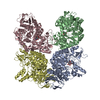
DNA BINDING PROTEIN / MutS / Mismatch sensor / ATPase
Function / homology
Function and homology information


mismatched DNA binding / ATP-dependent DNA damage sensor activity / mismatch repair / damaged DNA binding / ATP binding
Similarity search - Function
DNA repair protein MutS, domain I / MutS, DNA mismatch repair protein; Chain A, domain 3 / MutS, DNA mismatch repair protein; Chain A, domain 3 - #10 / DNA mismatch repair protein MutS / DNA mismatch repair protein MutS/MSH / DNA mismatch repair protein MutS-like, N-terminal / DNA mismatch repair protein MutS, connector domain / DNA mismatch repair protein MutS, clamp / DNA mismatch repair protein MutS, N-terminal / MutS, connector domain superfamily ...DNA repair protein MutS, domain I / MutS, DNA mismatch repair protein; Chain A, domain 3 / MutS, DNA mismatch repair protein; Chain A, domain 3 - #10 / DNA mismatch repair protein MutS / DNA mismatch repair protein MutS/MSH / DNA mismatch repair protein MutS-like, N-terminal / DNA mismatch repair protein MutS, connector domain / DNA mismatch repair protein MutS, clamp / DNA mismatch repair protein MutS, N-terminal / MutS, connector domain superfamily / MutS domain I / MutS domain II / MutS family domain IV / MutS domain III / DNA mismatch repair MutS family / DNA mismatch repair protein MutS, C-terminal / DNA mismatch repair protein MutS, core / DNA mismatch repair protein MutS, core domain superfamily / MutS domain V / DNA mismatch repair proteins mutS family signature. / DNA-binding domain of DNA mismatch repair MUTS family / ATPase domain of DNA mismatch repair MUTS family / MutS, DNA mismatch repair protein, domain I / P-loop containing nucleotide triphosphate hydrolases / P-loop containing nucleoside triphosphate hydrolase / Rossmann fold / Orthogonal Bundle / 3-Layer(aba) Sandwich / Mainly Alpha / Alpha Beta
Similarity search - Domain/homology
ADENOSINE-5'-DIPHOSPHATE / DNA mismatch repair protein MutS
Similarity search - Component
Biological speciesNeisseria gonorrhoeae (bacteria)
MethodX-RAY DIFFRACTION / SYNCHROTRON / MOLECULAR REPLACEMENT / Resolution: 2.97 Å
AuthorsNirwal, S. / Nair, D.T.
Funding support India, 1items
OrganizationGrant numberCountry
Department of BiotechnologyBT/PR882/BRB/10/935/2011 India
CitationJournal: Nucleic Acids Res. / Year: 2018
Title: Mechanism of formation of a toroid around DNA by the mismatch sensor protein.
Authors: Nirwal, S. / Kulkarni, D.S. / Sharma, A. / Rao, D.N. / Nair, D.T.
History
DepositionOct 12, 2017Deposition site: PDBJ / Processing site: PDBJ
Revision 1.0Nov 22, 2017Provider: repository / Type: Initial release
Revision 1.1Dec 13, 2017Group: Database references / Category: citation / Item: _citation.pdbx_database_id_PubMed / _citation.title
Revision 1.2Jan 24, 2018Group: Database references / Category: citation
Item: _citation.journal_volume / _citation.page_first ..._citation.journal_volume / _citation.page_first / _citation.page_last / _citation.year
Revision 1.3Sep 5, 2018Group: Data collection / Database references / Category: pdbx_related_exp_data_set
Revision 1.4Nov 22, 2023Group: Data collection / Database references / Refinement description
Category: chem_comp_atom / chem_comp_bond ...chem_comp_atom / chem_comp_bond / database_2 / pdbx_initial_refinement_model
Item: _database_2.pdbx_DOI / _database_2.pdbx_database_accession

-
Structure visualization

Structure viewerMolecule:
MolmilJmol/JSmol

Downloads & links

-
Assembly

Deposited unit
A: DNA mismatch repair protein MutS
B: DNA mismatch repair protein MutS
hetero molecules


Theoretical massNumber of molelcules
Total (without water)181,0544
Polymers180,2002
Non-polymers8542
Water2,144119
1


  • Idetical with deposited unit
  • defined by author&software
  • Evidence: SAXS
TypeNameSymmetry operationNumber
identity operation1_555x,y,z1
Buried area5200 Å2
ΔGint-37 kcal/mol
Surface area70040 Å2
MethodPISA
Unit cell
Length a, b, c (Å)89.940, 102.400, 236.170
Angle α, β, γ (deg.)90.00, 90.00, 90.00
Int Tables number18
Space group name H-MP21221

-
Components

#1: Protein DNA mismatch repair protein MutS /


Mass: 90099.852 Da / Num. of mol.: 2 / Fragment: UNP RESIDUES 1-814
Source method: isolated from a genetically manipulated source
Source: (gene. exp.) Neisseria gonorrhoeae (strain ATCC 700825 / FA 1090) (bacteria)
Strain: ATCC 700825 / FA 1090 / Gene: mutS, NGO1930 / Plasmid: PGEX6P1 / Production host: Escherichia coli (E. coli) / Strain (production host): C41DE3 / References: UniProt: Q5F5J4
#2: Chemical ChemComp-ADP / ADENOSINE-5'-DIPHOSPHATE / Adenosine diphosphate


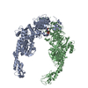
Mass: 427.201 Da / Num. of mol.: 2 / Source method: obtained synthetically / Formula: C10H15N5O10P2 / Feature type: SUBJECT OF INVESTIGATION / Comment: ADP, energy-carrying molecule*YM
#3: Water ChemComp-HOH / water / Water


Mass: 18.015 Da / Num. of mol.: 119 / Source method: isolated from a natural source / Formula: H2O

-
Experimental details

-
Experiment

ExperimentMethod: X-RAY DIFFRACTION / Number of used crystals: 1

-
Sample preparation

CrystalDensity Matthews: 3.19 Å3/Da / Density % sol: 61.43 % / Description: FLAT RECTANGLES
Crystal growTemperature: 300 K / Method: vapor diffusion, hanging drop / pH: 6.5
Details: 10-14% (W/V) PEG 4000, 0.1-0.2M MGCL2, PH 6.5, VAPOR DIFFUSION, HANGING DROP, TEMPERATURE 298K

-
Data collection

DiffractionMean temperature: 100 K
Diffraction sourceSource: SYNCHROTRON / Site: ESRF / Beamline: BM14 / Wavelength: 0.9537 Å
DetectorType: MARMOSAIC 225 mm CCD / Detector: CCD / Date: Oct 15, 2014
RadiationProtocol: SINGLE WAVELENGTH / Monochromatic (M) / Laue (L): M / Scattering type: x-ray
Radiation wavelengthWavelength: 0.9537 Å / Relative weight: 1
ReflectionResolution: 2.97→50 Å / Num. obs: 43285 / % possible obs: 98.7 % / Redundancy: 3.5 % / Rmerge(I) obs: 0.064 / Net I/σ(I): 8.6
Reflection shellResolution: 2.97→3.05 Å / Redundancy: 3.6 % / Rmerge(I) obs: 0.56 / Mean I/σ(I) obs: 2 / % possible all: 99.2

-
Processing

Software
NameVersionClassification
iMOSFLMdata reduction
SCALAdata scaling
PHASERphasing
REFMAC5.7.0032refinement
RefinementMethod to determine structure: MOLECULAR REPLACEMENT
Starting model: 1E3M
Resolution: 2.97→50 Å / Cor.coef. Fo:Fc: 0.923 / Cor.coef. Fo:Fc free: 0.904 / SU B: 42.556 / SU ML: 0.382 / Cross valid method: THROUGHOUT / ESU R Free: 0.429
RfactorNum. reflection% reflectionSelection details
Rfree0.269 2301 5 %RANDOM
Rwork0.246 ---
obs0.247 43285 99.4 %-
Solvent computationIon probe radii: 0.8 Å / Shrinkage radii: 0.8 Å / VDW probe radii: 1.2 Å
Displacement parametersBiso mean: 99.33 Å2
Baniso -1Baniso -2Baniso -3
1-3.63 Å20 Å20 Å2
2--0.57 Å20 Å2
3----4.2 Å2
Refinement stepCycle: LAST / Resolution: 2.97→50 Å
ProteinNucleic acidLigandSolventTotal
Num. atoms11810 0 54 119 11983
Refine LS restraints
Refine-IDTypeDev idealDev ideal targetNumber
X-RAY DIFFRACTIONr_bond_refined_d0.0060.01912088
X-RAY DIFFRACTIONr_bond_other_d
X-RAY DIFFRACTIONr_angle_refined_deg1.0841.96916400
X-RAY DIFFRACTIONr_angle_other_deg
X-RAY DIFFRACTIONr_dihedral_angle_1_deg6.14751533
X-RAY DIFFRACTIONr_dihedral_angle_2_deg38.63424.117532
X-RAY DIFFRACTIONr_dihedral_angle_3_deg18.812152041
X-RAY DIFFRACTIONr_dihedral_angle_4_deg11.6011579
X-RAY DIFFRACTIONr_chiral_restr0.0590.21895
X-RAY DIFFRACTIONr_gen_planes_refined0.0050.0219070
X-RAY DIFFRACTIONr_gen_planes_other
X-RAY DIFFRACTIONr_nbd_refined
X-RAY DIFFRACTIONr_nbd_other
X-RAY DIFFRACTIONr_nbtor_refined
X-RAY DIFFRACTIONr_nbtor_other
X-RAY DIFFRACTIONr_xyhbond_nbd_refined
X-RAY DIFFRACTIONr_xyhbond_nbd_other
X-RAY DIFFRACTIONr_metal_ion_refined
X-RAY DIFFRACTIONr_metal_ion_other
X-RAY DIFFRACTIONr_symmetry_vdw_refined
X-RAY DIFFRACTIONr_symmetry_vdw_other
X-RAY DIFFRACTIONr_symmetry_hbond_refined
X-RAY DIFFRACTIONr_symmetry_hbond_other
X-RAY DIFFRACTIONr_symmetry_metal_ion_refined
X-RAY DIFFRACTIONr_symmetry_metal_ion_other
X-RAY DIFFRACTIONr_mcbond_it4.5245.2976147
X-RAY DIFFRACTIONr_mcbond_other
X-RAY DIFFRACTIONr_mcangle_it7.47.9437675
X-RAY DIFFRACTIONr_mcangle_other
X-RAY DIFFRACTIONr_scbond_it4.895.5125941
X-RAY DIFFRACTIONr_scbond_other
X-RAY DIFFRACTIONr_scangle_it
X-RAY DIFFRACTIONr_scangle_other
X-RAY DIFFRACTIONr_long_range_B_refined14.26349.54750319
X-RAY DIFFRACTIONr_long_range_B_other
X-RAY DIFFRACTIONr_rigid_bond_restr
X-RAY DIFFRACTIONr_sphericity_free
X-RAY DIFFRACTIONr_sphericity_bonded
LS refinement shellResolution: 2.97→3.05 Å / Total num. of bins used: 20
RfactorNum. reflection% reflection
Rfree0.365 190 -
Rwork0.34 3115 -
obs--99.88 %
Refinement TLS params.

Method: refined / Refine-ID: X-RAY DIFFRACTION

IDL112)L122)L132)L222)L232)L332)S11 (Å °)S12 (Å °)S13 (Å °)S21 (Å °)S22 (Å °)S23 (Å °)S31 (Å °)S32 (Å °)S33 (Å °)T112)T122)T132)T222)T232)T332)Origin x (Å)Origin y (Å)Origin z (Å)
11.7732-0.3352-1.59560.11570.21262.24160.04060.0274-0.1029-0.11570.0279-0.0449-0.0134-0.0163-0.06840.3791-0.01940.0390.56170.00330.46343.8275-3.4131-11.8828
20.3484-0.21390.61471.0479-0.09471.8854-0.033-0.0551-0.0485-0.2771-0.0131-0.0335-0.1346-0.24790.04610.46660.00430.00410.5486-0.00380.424915.188911.376-34.6943
30.41530.31530.28212.34330.65270.32370.0791-0.08950.0308-0.8244-0.0724-0.2681-0.08820.0743-0.00670.76110.07460.18650.48870.05130.393836.2463-10.6219-53.3766
40.6875-0.2343-1.0340.27810.30542.5043-0.0772-0.2086-0.0153-0.02630.0282-0.0787-0.29070.30360.04910.3926-0.08280.0150.6023-0.00960.464740.80087.9137-23.3954
50.61670.848-1.60691.9532-1.49395.24820.0982-0.00650.03670.43370.0546-0.0094-0.3204-0.1214-0.15280.43950.0296-0.01740.6708-0.00910.302721.34985.818717.1759
61.81190.02092.13170.2729-0.47213.8307-0.06060.3348-0.15250.21110.17330.085-0.78230.267-0.11270.5677-0.0590.15690.3764-0.02960.537542.1903-48.9381-11.7609
70.8769-0.12910.45172.665-0.71571.684-0.0683-0.03380.0226-0.07710.26070.18510.09690.4559-0.19250.22040.02210.08470.44830.0470.586634.2192-61.805-37.0254
80.4059-0.35160.20363.0535-0.56410.24570.0864-0.0852-0.3303-0.97960.13170.8093-0.0346-0.0564-0.21820.81280.0503-0.29380.20630.0160.603915.4912-37.7884-56.3798
90.6541.05690.00421.8447-0.09752.66230.0074-0.12430.3330.1194-0.10370.89710.2476-0.18470.09630.10670.10510.27420.31680.17351.1757.4669-59.0286-29.1241
101.24850.38792.74091.41770.78026.03840.0958-0.15280.01130.3521-0.03370.14670.1406-0.4291-0.06220.42250.04820.43230.73990.24910.5322.034-58.733513.9298
Refinement TLS group
IDRefine-IDRefine TLS-IDAuth asym-IDAuth seq-ID
1X-RAY DIFFRACTION1A1 - 120
2X-RAY DIFFRACTION2A123 - 261
3X-RAY DIFFRACTION3A263 - 310
4X-RAY DIFFRACTION3A560 - 785
5X-RAY DIFFRACTION3A901
6X-RAY DIFFRACTION4A313 - 393
7X-RAY DIFFRACTION4A532 - 558
8X-RAY DIFFRACTION5A395 - 531
9X-RAY DIFFRACTION6B4 - 120
10X-RAY DIFFRACTION7B123 - 261
11X-RAY DIFFRACTION8B263 - 310
12X-RAY DIFFRACTION8B560 - 785
13X-RAY DIFFRACTION8B901
14X-RAY DIFFRACTION9B313 - 393
15X-RAY DIFFRACTION9B532 - 558
16X-RAY DIFFRACTION10B395 - 531

+
About Yorodumi

-
News

-
Feb 9, 2022. New format data for meta-information of EMDB entries

New format data for meta-information of EMDB entries

  • Version 3 of the EMDB header file is now the official format.
  • The previous official version 1.9 will be removed from the archive.

Related info.:EMDB header

External links:wwPDB to switch to version 3 of the EMDB data model

-
Aug 12, 2020. Covid-19 info

Covid-19 info

URL: https://pdbj.org/emnavi/covid19.php

New page: Covid-19 featured information page in EM Navigator.

Related info.:Covid-19 info / Mar 5, 2020. Novel coronavirus structure data

+
Mar 5, 2020. Novel coronavirus structure data

Novel coronavirus structure data

Related info.:Yorodumi Speices / Aug 12, 2020. Covid-19 info

External links:COVID-19 featured content - PDBj / Molecule of the Month (242):Coronavirus Proteases

+
Jan 31, 2019. EMDB accession codes are about to change! (news from PDBe EMDB page)

EMDB accession codes are about to change! (news from PDBe EMDB page)

  • The allocation of 4 digits for EMDB accession codes will soon come to an end. Whilst these codes will remain in use, new EMDB accession codes will include an additional digit and will expand incrementally as the available range of codes is exhausted. The current 4-digit format prefixed with “EMD-” (i.e. EMD-XXXX) will advance to a 5-digit format (i.e. EMD-XXXXX), and so on. It is currently estimated that the 4-digit codes will be depleted around Spring 2019, at which point the 5-digit format will come into force.
  • The EM Navigator/Yorodumi systems omit the EMD- prefix.

Related info.:Q: What is EMD? / ID/Accession-code notation in Yorodumi/EM Navigator

External links:EMDB Accession Codes are Changing Soon! / Contact to PDBj

+
Jul 12, 2017. Major update of PDB

Major update of PDB

  • wwPDB released updated PDB data conforming to the new PDBx/mmCIF dictionary.
  • This is a major update changing the version number from 4 to 5, and with Remediation, in which all the entries are updated.
  • In this update, many items about electron microscopy experimental information are reorganized (e.g. em_software).
  • Now, EM Navigator and Yorodumi are based on the updated data.

External links:wwPDB Remediation / Enriched Model Files Conforming to OneDep Data Standards Now Available in the PDB FTP Archive

-
Yorodumi

Thousand views of thousand structures

  • Yorodumi is a browser for structure data from EMDB, PDB, SASBDB, etc.
  • This page is also the successor to EM Navigator detail page, and also detail information page/front-end page for Omokage search.
  • The word "yorodu" (or yorozu) is an old Japanese word meaning "ten thousand". "mi" (miru) is to see.

Related info.:EMDB / PDB / SASBDB / Comparison of 3 databanks / Yorodumi Search / Aug 31, 2016. New EM Navigator & Yorodumi / Yorodumi Papers / Jmol/JSmol / Function and homology information / Changes in new EM Navigator and Yorodumi

Read more