 Movie
Movie Controller


+ Open data

- Basic information
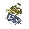
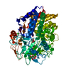
| Entry | Database: PDB / ID: 5yj6 | ||||||
|---|---|---|---|---|---|---|---|
| Title | The exoglucanase CelS from Clostridium thermocellum | ||||||
 Components Components | Dockerin type I repeat-containing protein | ||||||
 Keywords Keywords |  HYDROLASE / activity / cellulosome / exocellulase / substrate specificity HYDROLASE / activity / cellulosome / exocellulase / substrate specificity | ||||||
| Function / homology |  Function and homology information Function and homology information | ||||||
| Biological species |  Ruminiclostridium thermocellum DSM 1313 (bacteria) Ruminiclostridium thermocellum DSM 1313 (bacteria) | ||||||
| Method | X-RAY DIFFRACTION / SYNCHROTRON / MOLECULAR REPLACEMENT / Resolution: 1.43 Å | ||||||
 Authors Authors | Liu, Y.J. / Liu, S.Y. / Dong, S. / Li, R.M. / Feng, Y.G. / Cui, Q. | ||||||
 Citation Citation | Journal: Biotechnol Biofuels / Year: 2018 Title: Determination of the native features of the exoglucanase Cel48S from Clostridium thermocellum Authors: Liu, Y.J. / Liu, S. / Dong, S. / Li, R. / Feng, Y. / Cui, Q. | ||||||
| History |
|
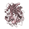
- Structure visualization
| Structure viewer | Molecule: MolmilJmol/JSmol |
|---|
- Downloads & links
-Download
| PDBx/mmCIF format | 5yj6.cif.gz | 389.3 KB | Display | PDBx/mmCIF format |
|---|---|---|---|---|
| PDB format | pdb5yj6.ent.gz | 317.6 KB | Display | PDB format |
| PDBx/mmJSON format | 5yj6.json.gz | Tree view | PDBx/mmJSON format | |
| Others |  Other downloads Other downloads |
-Validation report
| Arichive directory | https://data.pdbj.org/pub/pdb/validation_reports/yj/5yj6ftp://data.pdbj.org/pub/pdb/validation_reports/yj/5yj6 | HTTPS FTP |
|---|
-Related structure data
| Related structure data |  1l2aS S: Starting model for refinement |
|---|---|
| Similar structure data |


-Links
 PDBj
PDBj
- Assembly
| Deposited unit | 
| ||||||||||||
|---|---|---|---|---|---|---|---|---|---|---|---|---|---|
| 1 |
| ||||||||||||
| Unit cell |
| ||||||||||||
| Components on special symmetry positions |
|
-Components
| #1: Protein | Mass: 74347.039 Da / Num. of mol.: 1 / Fragment: UNP residues 31-666 Source method: isolated from a genetically manipulated source Source: (gene. exp.) Ruminiclostridium thermocellum DSM 1313 (bacteria)Gene: SAMN04515622_0728 / Production host: Escherichia coli (E. coli) / References: UniProt: A0A286AKY4, UniProt: C7ED31*PLUS |
|---|---|
| #2: Chemical | ChemComp-33O / 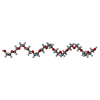  Mass: 590.699 Da / Num. of mol.: 1 / Source method: obtained synthetically / Formula: C26H54O14 Mass: 590.699 Da / Num. of mol.: 1 / Source method: obtained synthetically / Formula: C26H54O14 |
| #3: Water | ChemComp-HOH / Water Mass: 18.015 Da / Num. of mol.: 825 / Source method: isolated from a natural source / Formula: H2O Mass: 18.015 Da / Num. of mol.: 825 / Source method: isolated from a natural source / Formula: H2O |
-Experimental details
-Experiment
| Experiment | Method: X-RAY DIFFRACTION / Number of used crystals: 1 |
|---|
- Sample preparation
| Crystal | Density Matthews: 1.77 Å3/Da / Density % sol: 36.07 % |
|---|---|
| Crystal grow | Temperature: 291 K / Method: vapor diffusion, hanging drop / pH: 7.2 / Details: 200mM sodium formate, 20% PEG3350 |
-Data collection
| Diffraction | Mean temperature: 100 K | ||||||||||||||||||||||||||||||
|---|---|---|---|---|---|---|---|---|---|---|---|---|---|---|---|---|---|---|---|---|---|---|---|---|---|---|---|---|---|---|---|
| Diffraction source | Source: SYNCHROTRON / Site: SSRF  / Beamline: BL17U1 / Wavelength: 0.979 Å / Beamline: BL17U1 / Wavelength: 0.979 Å | ||||||||||||||||||||||||||||||
| Detector | Type: ADSC QUANTUM 315r / Detector: CCD / Date: Dec 21, 2016 | ||||||||||||||||||||||||||||||
| Radiation | Protocol: SINGLE WAVELENGTH / Monochromatic (M) / Laue (L): M / Scattering type: x-ray | ||||||||||||||||||||||||||||||
| Radiation wavelength | Wavelength: 0.979 Å / Relative weight: 1 | ||||||||||||||||||||||||||||||
| Reflection | Resolution: 1.43→101.07 Å / Num. obs: 100821 / % possible obs: 94.9 % / Redundancy: 7.3 % / CC1/2: 0.999 / Rmerge(I) obs: 0.075 / Rpim(I) all: 0.03 / Rrim(I) all: 0.08 / Net I/σ(I): 16 / Num. measured all: 737296 / Scaling rejects: 0 | ||||||||||||||||||||||||||||||
| Reflection shell | Diffraction-ID: 1
|
- Processing
| Software |
| |||||||||||||||||||||||||||||||||||||||||||||||||||||||||||||||||||||||||||||||||||||||||||||||||||||||||||||||||||||||||||||
|---|---|---|---|---|---|---|---|---|---|---|---|---|---|---|---|---|---|---|---|---|---|---|---|---|---|---|---|---|---|---|---|---|---|---|---|---|---|---|---|---|---|---|---|---|---|---|---|---|---|---|---|---|---|---|---|---|---|---|---|---|---|---|---|---|---|---|---|---|---|---|---|---|---|---|---|---|---|---|---|---|---|---|---|---|---|---|---|---|---|---|---|---|---|---|---|---|---|---|---|---|---|---|---|---|---|---|---|---|---|---|---|---|---|---|---|---|---|---|---|---|---|---|---|---|---|---|
| Refinement | Method to determine structure: MOLECULAR REPLACEMENT Starting model: 1L2A Resolution: 1.43→38.373 Å / SU ML: 0.13 / Cross valid method: FREE R-VALUE / σ(F): 1.35 / Phase error: 20.8 / Stereochemistry target values: ML
| |||||||||||||||||||||||||||||||||||||||||||||||||||||||||||||||||||||||||||||||||||||||||||||||||||||||||||||||||||||||||||||
| Solvent computation | Shrinkage radii: 0.9 Å / VDW probe radii: 1.11 Å / Solvent model: FLAT BULK SOLVENT MODEL | |||||||||||||||||||||||||||||||||||||||||||||||||||||||||||||||||||||||||||||||||||||||||||||||||||||||||||||||||||||||||||||
| Displacement parameters | Biso max: 97.97 Å2 / Biso mean: 23.3299 Å2 / Biso min: 11.05 Å2 | |||||||||||||||||||||||||||||||||||||||||||||||||||||||||||||||||||||||||||||||||||||||||||||||||||||||||||||||||||||||||||||
| Refinement step | Cycle: final / Resolution: 1.43→38.373 Å
| |||||||||||||||||||||||||||||||||||||||||||||||||||||||||||||||||||||||||||||||||||||||||||||||||||||||||||||||||||||||||||||
| Refinement TLS params. | Method: refined / Refine-ID: X-RAY DIFFRACTION
| |||||||||||||||||||||||||||||||||||||||||||||||||||||||||||||||||||||||||||||||||||||||||||||||||||||||||||||||||||||||||||||
| Refinement TLS group |
|
