 Movie
Movie Controller

[English] 日本語
 Yorodumi
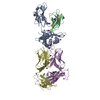
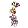
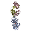
- PDB-5yhl: Crystal structure of the human prostaglandin E receptor EP4 in co... -
+ Open data

- Basic information
| Entry | Database: PDB / ID: 5yhl | |||||||||
|---|---|---|---|---|---|---|---|---|---|---|
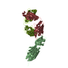
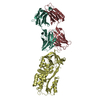
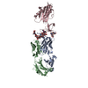
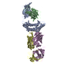
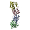
| Title | Crystal structure of the human prostaglandin E receptor EP4 in complex with Fab and an antagonist Br-derivative | |||||||||
 Components Components |
| |||||||||
 Keywords Keywords |  SIGNALING PROTEIN/IMMUNE SYSTEM / G-protein coupled receptor / lipid mediator / functional antibody / SIGNALING PROTEIN-IMMUNE SYSTEM complex SIGNALING PROTEIN/IMMUNE SYSTEM / G-protein coupled receptor / lipid mediator / functional antibody / SIGNALING PROTEIN-IMMUNE SYSTEM complex | |||||||||
| Function / homology |  Function and homology information Function and homology informationnegative regulation of eosinophil extravasation / prostaglandin E receptor activity / Prostanoid ligand receptors / negative regulation of integrin activation / response to nematode / T-helper cell differentiation / regulation of stress fiber assembly / negative regulation of cytokine production / regulation of ossification / response to mechanical stimulus ...negative regulation of eosinophil extravasation / prostaglandin E receptor activity / Prostanoid ligand receptors / negative regulation of integrin activation / response to nematode / T-helper cell differentiation / regulation of stress fiber assembly / negative regulation of cytokine production / regulation of ossification / response to mechanical stimulus / JNK cascade / ERK1 and ERK2 cascade / positive regulation of cytokine production / bone development / adenylate cyclase-modulating G protein-coupled receptor signaling pathway / adenylate cyclase-activating G protein-coupled receptor signaling pathway / negative regulation of inflammatory response / positive regulation of inflammatory response / cellular response to mechanical stimulus / cellular response to prostaglandin E stimulus / positive regulation of cytosolic calcium ion concentration / G alpha (s) signalling events / response to lipopolysaccharide / immune response / inflammatory response / membrane / plasma membraneSimilarity search - Function | |||||||||
| Biological species |  Homo sapiens (human) Homo sapiens (human) Mus musculus (house mouse) Mus musculus (house mouse) | |||||||||
| Method | X-RAY DIFFRACTION / SYNCHROTRON / MOLECULAR REPLACEMENT / Resolution: 4.2 Å | |||||||||
 Authors Authors | Toyoda, Y. / Morimoto, K. / Suno, R. / Horita, S. / Iwata, S. / Kobayashi, T. | |||||||||
| Funding support |  Japan, 2items Japan, 2items
| |||||||||
 Citation Citation | Journal: Nat. Chem. Biol. / Year: 2019 Title: Ligand binding to human prostaglandin E receptor EP4at the lipid-bilayer interface. Authors: Toyoda, Y. / Morimoto, K. / Suno, R. / Horita, S. / Yamashita, K. / Hirata, K. / Sekiguchi, Y. / Yasuda, S. / Shiroishi, M. / Shimizu, T. / Urushibata, Y. / Kajiwara, Y. / Inazumi, T. / ...Authors: Toyoda, Y. / Morimoto, K. / Suno, R. / Horita, S. / Yamashita, K. / Hirata, K. / Sekiguchi, Y. / Yasuda, S. / Shiroishi, M. / Shimizu, T. / Urushibata, Y. / Kajiwara, Y. / Inazumi, T. / Hotta, Y. / Asada, H. / Nakane, T. / Shiimura, Y. / Nakagita, T. / Tsuge, K. / Yoshida, S. / Kuribara, T. / Hosoya, T. / Sugimoto, Y. / Nomura, N. / Sato, M. / Hirokawa, T. / Kinoshita, M. / Murata, T. / Takayama, K. / Yamamoto, M. / Narumiya, S. / Iwata, S. / Kobayashi, T. | |||||||||
| History |
|
- Structure visualization
| Structure viewer | Molecule: MolmilJmol/JSmol |
|---|
- Downloads & links
-Download
| PDBx/mmCIF format | 5yhl.cif.gz | 158.4 KB | Display | PDBx/mmCIF format |
|---|---|---|---|---|
| PDB format | pdb5yhl.ent.gz | 120.2 KB | Display | PDB format |
| PDBx/mmJSON format | 5yhl.json.gz | Tree view | PDBx/mmJSON format | |
| Others |  Other downloads Other downloads |
-Validation report
| Arichive directory | https://data.pdbj.org/pub/pdb/validation_reports/yh/5yhlftp://data.pdbj.org/pub/pdb/validation_reports/yh/5yhl | HTTPS FTP |
|---|
-Related structure data
| Related structure data |  5yfiC 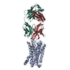 5ywyC  5wv2 S: Starting model for refinement C: citing same article ( |
|---|---|
| Similar structure data | |
| Experimental dataset #1 | Data reference: 10.5281/zenodo.1173791 / Data set type: diffraction image data / Details: Raw data |


-Links
 PDBj
PDBj


- Assembly
| Deposited unit | 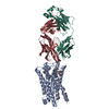
| ||||||||
|---|---|---|---|---|---|---|---|---|---|
| 1 |
| ||||||||
| Unit cell |
|
-Components
| #1: Protein | Mass: 37063.547 Da / Num. of mol.: 1 / Fragment: UNP RESIDUES 4-217, 260-366 / Mutation: N7Q, A62L, G106R, N177Q, 218-259 deletion Source method: isolated from a genetically manipulated source Source: (gene. exp.) Homo sapiens (human) / Gene: PTGER4, PTGER2 / Production host:   Spodoptera frugiperda (fall armyworm) / References: UniProt: P35408 Spodoptera frugiperda (fall armyworm) / References: UniProt: P35408 |
|---|---|
| #2: Antibody | Mass: 27574.092 Da / Num. of mol.: 1 Source method: isolated from a genetically manipulated source Source: (gene. exp.) Mus musculus (house mouse) / Production host: hybrid (others) |
| #3: Antibody | Mass: 26614.857 Da / Num. of mol.: 1 Source method: isolated from a genetically manipulated source Source: (gene. exp.) Mus musculus (house mouse) / Production host: hybrid (others) |
| #4: Chemical | ChemComp-8VL / 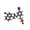  Mass: 465.339 Da / Num. of mol.: 1 / Source method: obtained synthetically / Formula: C24H21BrN2O3 Mass: 465.339 Da / Num. of mol.: 1 / Source method: obtained synthetically / Formula: C24H21BrN2O3 |
-Experimental details
-Experiment
| Experiment | Method: X-RAY DIFFRACTION / Number of used crystals: 1 |
|---|
- Sample preparation
| Crystal | Density Matthews: 4.44 Å3/Da / Density % sol: 72.31 % Description: The entry contains friedel pairs in F_plus/minus columns and I_plus/minus columns |
|---|---|
| Crystal grow | Temperature: 293 K / Method: lipidic cubic phase Details: 24-31% PEG 300, 150 mM potassium sulfate, 100 mM MES (pH 5.5-6.5), 1% 1,2,3-heptanetriol, 0.2 mM ONO-AE3-208-Br, 2% DMSO PH range: 5.5-6.5 |
-Data collection
| Diffraction | Mean temperature: 100 K | ||||||||||||||||||||||||||||||||||||||||||||||||||||||||||||||||||||||||||||||||
|---|---|---|---|---|---|---|---|---|---|---|---|---|---|---|---|---|---|---|---|---|---|---|---|---|---|---|---|---|---|---|---|---|---|---|---|---|---|---|---|---|---|---|---|---|---|---|---|---|---|---|---|---|---|---|---|---|---|---|---|---|---|---|---|---|---|---|---|---|---|---|---|---|---|---|---|---|---|---|---|---|---|
| Diffraction source | Source: SYNCHROTRON / Site: SPring-8 / Beamline: BL32XU / Wavelength: 0.9 Å | ||||||||||||||||||||||||||||||||||||||||||||||||||||||||||||||||||||||||||||||||
| Detector | Type: DECTRIS EIGER X 9M / Detector: PIXEL / Date: May 16, 2016 | ||||||||||||||||||||||||||||||||||||||||||||||||||||||||||||||||||||||||||||||||
| Radiation | Monochromator: Si(111) / Protocol: SINGLE WAVELENGTH / Monochromatic (M) / Laue (L): M / Scattering type: x-ray | ||||||||||||||||||||||||||||||||||||||||||||||||||||||||||||||||||||||||||||||||
| Radiation wavelength | Wavelength: 0.9 Å / Relative weight: 1 | ||||||||||||||||||||||||||||||||||||||||||||||||||||||||||||||||||||||||||||||||
| Reflection | Resolution: 4.2→49.339 Å / Num. obs: 22914 / % possible obs: 99.9 % / Observed criterion σ(I): -3 / Redundancy: 15.076 % / Biso Wilson estimate: 65.9 Å2 / CC1/2: 0.982 / Rmerge(I) obs: 0.0716 / Rrim(I) all: 0.741 / Χ2: 1.446 / Net I/σ(I): 4.31 | ||||||||||||||||||||||||||||||||||||||||||||||||||||||||||||||||||||||||||||||||
| Reflection shell | Diffraction-ID: 1
|
- Processing
| Software |
| |||||||||||||||||||||||||||||||||||||||||||||||||||||||||||||||
|---|---|---|---|---|---|---|---|---|---|---|---|---|---|---|---|---|---|---|---|---|---|---|---|---|---|---|---|---|---|---|---|---|---|---|---|---|---|---|---|---|---|---|---|---|---|---|---|---|---|---|---|---|---|---|---|---|---|---|---|---|---|---|---|---|
| Refinement | Method to determine structure: MOLECULAR REPLACEMENT Starting model: 5WV2 5wv2 Resolution: 4.2→49.339 Å / SU ML: 0.85 / Cross valid method: FREE R-VALUE / σ(F): 1.33 / Phase error: 34.28 Details: The entry contains friedel pairs in F_plus/minus columns and I_plus/minus columns
| |||||||||||||||||||||||||||||||||||||||||||||||||||||||||||||||
| Solvent computation | Shrinkage radii: 0.9 Å / VDW probe radii: 1.11 Å | |||||||||||||||||||||||||||||||||||||||||||||||||||||||||||||||
| Displacement parameters | Biso max: 266.01 Å2 / Biso mean: 134.3258 Å2 / Biso min: 76.03 Å2 | |||||||||||||||||||||||||||||||||||||||||||||||||||||||||||||||
| Refinement step | Cycle: final / Resolution: 4.2→49.339 Å
| |||||||||||||||||||||||||||||||||||||||||||||||||||||||||||||||
| Refine LS restraints |
| |||||||||||||||||||||||||||||||||||||||||||||||||||||||||||||||
| LS refinement shell | Refine-ID: X-RAY DIFFRACTION / Rfactor Rfree error: 0 / Total num. of bins used: 8
|
