 Movie
Movie Controller

[English] 日本語
 Yorodumi
- PDB-5yd5: Crystal structure of the scFv antibody 4B08 with epitope peptide ... -
+ Open data

- Basic information
| Entry | Database: PDB / ID: 5yd5 | |||||||||
|---|---|---|---|---|---|---|---|---|---|---|
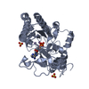
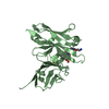
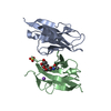
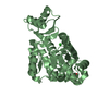
| Title | Crystal structure of the scFv antibody 4B08 with epitope peptide (mutation N3A) | |||||||||
 Components Components |
| |||||||||
 Keywords Keywords |  IMMUNE SYSTEM / Antibody / Biomolecular recognition / MD simulations / Thermodynamics IMMUNE SYSTEM / Antibody / Biomolecular recognition / MD simulations / Thermodynamics | |||||||||
| Function / homology |  Function and homology information Function and homology informationchemokine (C-C motif) ligand 5 binding / negative regulation of macrophage apoptotic process / signaling / chemokine receptor activity / C-C chemokine receptor activity / C-C chemokine binding / phosphatidylinositol phospholipase C activity / response to cholesterol / Chemokine receptors bind chemokines / release of sequestered calcium ion into cytosol by sarcoplasmic reticulum ...chemokine (C-C motif) ligand 5 binding / negative regulation of macrophage apoptotic process / signaling / chemokine receptor activity / C-C chemokine receptor activity / C-C chemokine binding / phosphatidylinositol phospholipase C activity / response to cholesterol / Chemokine receptors bind chemokines / release of sequestered calcium ion into cytosol by sarcoplasmic reticulum / dendritic cell chemotaxis / immunoglobulin complex / Interleukin-10 signaling / Binding and entry of HIV virion / cellular defense response / coreceptor activity / antigen binding / cell chemotaxis / calcium-mediated signaling / chemotaxis / calcium ion transport / MAPK cascade / virus receptor activity / cell-cell signaling / actin binding / G alpha (i) signalling events / positive regulation of cytosolic calcium ion concentration / cellular response to lipopolysaccharide / adaptive immune response / cell surface receptor signaling pathway / endosome / immune response / inflammatory response / G protein-coupled receptor signaling pathway / external side of plasma membrane / cell surface / extracellular space / identical protein binding / plasma membrane / cytoplasmSimilarity search - Function | |||||||||
| Biological species |  Mus musculus (house mouse) Mus musculus (house mouse) Homo sapiens (human) Homo sapiens (human) | |||||||||
| Method | X-RAY DIFFRACTION / SYNCHROTRON / MOLECULAR REPLACEMENT / Resolution: 1.96 Å | |||||||||
 Authors Authors | Caaveiro, J.M.M. / Miyanabe, K. / Tsumoto, K. | |||||||||
| Funding support |  Japan, 2items Japan, 2items
| |||||||||
 Citation Citation | Journal: J. Biochem. / Year: 2018 Title: Intramolecular H-bonds govern the recognition of a flexible peptide by an antibody Authors: Miyanabe, K. / Akiba, H. / Kuroda, D. / Nakakido, M. / Kusano-Arai, O. / Iwanari, H. / Hamakubo, T. / Caaveiro, J.M.M. / Tsumoto, K. | |||||||||
| History |
|
- Structure visualization
| Structure viewer | Molecule: MolmilJmol/JSmol |
|---|
- Downloads & links
-Download
| PDBx/mmCIF format | 5yd5.cif.gz | 116.9 KB | Display | PDBx/mmCIF format |
|---|---|---|---|---|
| PDB format | pdb5yd5.ent.gz | 89.8 KB | Display | PDB format |
| PDBx/mmJSON format | 5yd5.json.gz | Tree view | PDBx/mmJSON format | |
| Others |  Other downloads Other downloads |
-Validation report
| Arichive directory | https://data.pdbj.org/pub/pdb/validation_reports/yd/5yd5ftp://data.pdbj.org/pub/pdb/validation_reports/yd/5yd5 | HTTPS FTP |
|---|
-Related structure data
| Related structure data |  5yd3SC  5yd4C S: Starting model for refinement C: citing same article ( |
|---|---|
| Similar structure data |


-Links
 PDBj
PDBj


- Assembly
| Deposited unit | 
| ||||||||
|---|---|---|---|---|---|---|---|---|---|
| 1 | 
| ||||||||
| 2 | 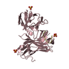
| ||||||||
| Unit cell |
| ||||||||
| Components on special symmetry positions |
|
-Components
| #1: Antibody | Mass: 26748.613 Da / Num. of mol.: 2 Source method: isolated from a genetically manipulated source Source: (gene. exp.) Mus musculus (house mouse) / Production host:  Escherichia coli BL21 (bacteria) / Strain (production host): BL21 / Variant (production host): DE3 / References: UniProt: P01630*PLUS Escherichia coli BL21 (bacteria) / Strain (production host): BL21 / Variant (production host): DE3 / References: UniProt: P01630*PLUS#2: Protein/peptide | Mass: 1058.096 Da / Num. of mol.: 2 / Source method: obtained synthetically / Source: (synth.) Homo sapiens (human) / References: UniProt: P51681*PLUS#3: Chemical | ChemComp-SO4 / Sulfate  Mass: 96.063 Da / Num. of mol.: 8 / Source method: obtained synthetically / Formula: SO4 Mass: 96.063 Da / Num. of mol.: 8 / Source method: obtained synthetically / Formula: SO4#4: Water | ChemComp-HOH / | Water Mass: 18.015 Da / Num. of mol.: 359 / Source method: isolated from a natural source / Formula: H2O Mass: 18.015 Da / Num. of mol.: 359 / Source method: isolated from a natural source / Formula: H2OCompound details | dimeric means one chain of antibody and one chain of peptide A + B or C + D | |
|---|
-Experimental details
-Experiment
| Experiment | Method: X-RAY DIFFRACTION / Number of used crystals: 1 |
|---|
- Sample preparation
| Crystal | Density Matthews: 2.59 Å3/Da / Density % sol: 52.53 % |
|---|---|
| Crystal grow | Temperature: 293.15 K / Method: vapor diffusion, hanging drop / Details: 100 mM BIS-TRIS 1.8 M ammonium sulphate |
-Data collection
| Diffraction | Mean temperature: 100 K |
|---|---|
| Diffraction source | Source: SYNCHROTRON / Site: Photon Factory / Beamline: AR-NE3A / Wavelength: 1 Å |
| Detector | Type: ADSC QUANTUM 270 / Detector: CCD / Date: May 18, 2015 |
| Radiation | Protocol: SINGLE WAVELENGTH / Monochromatic (M) / Laue (L): M / Scattering type: x-ray |
| Radiation wavelength | Wavelength: 1 Å / Relative weight: 1 |
| Reflection | Resolution: 1.96→37.3 Å / Num. obs: 40361 / % possible obs: 95.6 % / Redundancy: 7.7 % / CC1/2: 0.997 / Rmerge(I) obs: 0.129 / Rpim(I) all: 0.065 / Net I/σ(I): 10 |
| Reflection shell | Resolution: 1.96→2.01 Å / Redundancy: 6.2 % / Rmerge(I) obs: 0.425 / Mean I/σ(I) obs: 3.7 / Num. unique obs: 2555 / CC1/2: 0.939 / Rpim(I) all: 0.174 / % possible all: 87.1 |
- Processing
| Software |
| ||||||||||||||||||||||||||||||||||||||||||||||||||||||||||||||||||||||||||||||||||||||||||||||||||||||||||||||||||||||||||||||||||||||||||||||||||||||||||||||||||||||||||||||||||||||
|---|---|---|---|---|---|---|---|---|---|---|---|---|---|---|---|---|---|---|---|---|---|---|---|---|---|---|---|---|---|---|---|---|---|---|---|---|---|---|---|---|---|---|---|---|---|---|---|---|---|---|---|---|---|---|---|---|---|---|---|---|---|---|---|---|---|---|---|---|---|---|---|---|---|---|---|---|---|---|---|---|---|---|---|---|---|---|---|---|---|---|---|---|---|---|---|---|---|---|---|---|---|---|---|---|---|---|---|---|---|---|---|---|---|---|---|---|---|---|---|---|---|---|---|---|---|---|---|---|---|---|---|---|---|---|---|---|---|---|---|---|---|---|---|---|---|---|---|---|---|---|---|---|---|---|---|---|---|---|---|---|---|---|---|---|---|---|---|---|---|---|---|---|---|---|---|---|---|---|---|---|---|---|---|
| Refinement | Method to determine structure: MOLECULAR REPLACEMENT Starting model: 5YD3 Resolution: 1.96→37.3 Å / Cor.coef. Fo:Fc: 0.95 / Cor.coef. Fo:Fc free: 0.915 / SU B: 2.994 / SU ML: 0.088 / Cross valid method: THROUGHOUT / ESU R: 0.039 / ESU R Free: 0.037 / Details: HYDROGENS HAVE BEEN ADDED IN THE RIDING POSITIONS
| ||||||||||||||||||||||||||||||||||||||||||||||||||||||||||||||||||||||||||||||||||||||||||||||||||||||||||||||||||||||||||||||||||||||||||||||||||||||||||||||||||||||||||||||||||||||
| Solvent computation | Ion probe radii: 0.8 Å / Shrinkage radii: 0.8 Å / VDW probe radii: 1.2 Å | ||||||||||||||||||||||||||||||||||||||||||||||||||||||||||||||||||||||||||||||||||||||||||||||||||||||||||||||||||||||||||||||||||||||||||||||||||||||||||||||||||||||||||||||||||||||
| Displacement parameters | Biso mean: 16.291 Å2
| ||||||||||||||||||||||||||||||||||||||||||||||||||||||||||||||||||||||||||||||||||||||||||||||||||||||||||||||||||||||||||||||||||||||||||||||||||||||||||||||||||||||||||||||||||||||
| Refinement step | Cycle: 1 / Resolution: 1.96→37.3 Å
| ||||||||||||||||||||||||||||||||||||||||||||||||||||||||||||||||||||||||||||||||||||||||||||||||||||||||||||||||||||||||||||||||||||||||||||||||||||||||||||||||||||||||||||||||||||||
| Refine LS restraints |
|