 Movie
Movie Controller

[English] 日本語
 Yorodumi
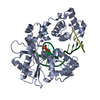
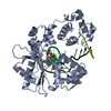
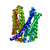
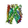
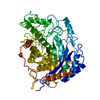
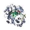
- PDB-5yck: Crystal structure of a MATE family protein derived from Camelina ... -
+ Open data

- Basic information
| Entry | Database: PDB / ID: 5yck | ||||||
|---|---|---|---|---|---|---|---|
| Title | Crystal structure of a MATE family protein derived from Camelina sativa at 2.3 angstrom | ||||||
 Components Components | multi drug efflux transporter | ||||||
 Keywords Keywords |  TRANSPORT PROTEIN / membrane protein / multi drug resistance / transporter TRANSPORT PROTEIN / membrane protein / multi drug resistance / transporter | ||||||
| Function / homology | Multi antimicrobial extrusion protein / MatE / antiporter activity / xenobiotic transmembrane transporter activity / membrane => GO:0016020 / (2R)-2,3-dihydroxypropyl (9Z)-octadec-9-enoate / RUBIDIUM ION / Protein DETOXIFICATION Function and homology information Function and homology information | ||||||
| Biological species |  Camelina sativa (false flax) Camelina sativa (false flax) | ||||||
| Method | X-RAY DIFFRACTION / SYNCHROTRON / MOLECULAR REPLACEMENT / molecular replacement / Resolution: 2.3 Å | ||||||
 Authors Authors | Tanaka, Y. / Tsukazaki, T. / Iwaki, S. | ||||||
 Citation Citation | Journal: Structure / Year: 2017 Title: Crystal Structure of a Plant Multidrug and Toxic Compound Extrusion Family Protein Authors: Tanaka, Y. / Iwaki, S. / Tsukazaki, T. | ||||||
| History |
|
- Structure visualization
| Structure viewer | Molecule: MolmilJmol/JSmol |
|---|
- Downloads & links
-Download
| PDBx/mmCIF format | 5yck.cif.gz | 105.6 KB | Display | PDBx/mmCIF format |
|---|---|---|---|---|
| PDB format | pdb5yck.ent.gz | 78 KB | Display | PDB format |
| PDBx/mmJSON format | 5yck.json.gz | Tree view | PDBx/mmJSON format | |
| Others |  Other downloads Other downloads |
-Validation report
| Arichive directory | https://data.pdbj.org/pub/pdb/validation_reports/yc/5yckftp://data.pdbj.org/pub/pdb/validation_reports/yc/5yck | HTTPS FTP |
|---|
-Related structure data
| Related structure data |  5xjjSC S: Starting model for refinement C: citing same article ( |
|---|---|
| Similar structure data |

-Links
 PDBj
PDBj


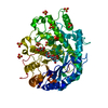
- Assembly
| Deposited unit | 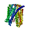
| ||||||||
|---|---|---|---|---|---|---|---|---|---|
| 1 |
| ||||||||
| Unit cell |
|
-Components
| #1: Protein | Mass: 51426.637 Da / Num. of mol.: 1 Source method: isolated from a genetically manipulated source Source: (gene. exp.) Camelina sativa (false flax) / Gene: false flax / Plasmid: pPic9k / Production host:  Komagataella pastoris (fungus) / References: UniProt: R0G998*PLUS Komagataella pastoris (fungus) / References: UniProt: R0G998*PLUS | ||||||
|---|---|---|---|---|---|---|---|
| #2: Chemical | Rubidium  Mass: 85.468 Da / Num. of mol.: 2 / Source method: obtained synthetically / Formula: Rb Mass: 85.468 Da / Num. of mol.: 2 / Source method: obtained synthetically / Formula: Rb#3: Chemical |   Mass: 356.540 Da / Num. of mol.: 3 / Source method: obtained synthetically / Formula: C21H40O4 Mass: 356.540 Da / Num. of mol.: 3 / Source method: obtained synthetically / Formula: C21H40O4#4: Water | ChemComp-HOH / | Water Mass: 18.015 Da / Num. of mol.: 84 / Source method: isolated from a natural source / Formula: H2O Mass: 18.015 Da / Num. of mol.: 84 / Source method: isolated from a natural source / Formula: H2OSequence details | AUTHORS USED THE NCBI Reference Sequence: XP_010514235.1. | |
-Experimental details
-Experiment
| Experiment | Method: X-RAY DIFFRACTION / Number of used crystals: 1 |
|---|
- Sample preparation
| Crystal | Density Matthews: 2.5 Å3/Da / Density % sol: 50.89 % |
|---|---|
| Crystal grow | Temperature: 298 K / Method: lipidic cubic phase / pH: 7.1 Details: 33% PEG300, 200mM (NH4)2SO4, 100mM Tris, 100mM RbCl |
-Data collection
| Diffraction | Mean temperature: 100 K | ||||||||||||||||||||||||||||||||||||||||||||||||||||||||||||||||||||||||||||||||
|---|---|---|---|---|---|---|---|---|---|---|---|---|---|---|---|---|---|---|---|---|---|---|---|---|---|---|---|---|---|---|---|---|---|---|---|---|---|---|---|---|---|---|---|---|---|---|---|---|---|---|---|---|---|---|---|---|---|---|---|---|---|---|---|---|---|---|---|---|---|---|---|---|---|---|---|---|---|---|---|---|---|
| Diffraction source | Source: SYNCHROTRON / Site: SPring-8  / Beamline: BL32XU / Wavelength: 1 Å / Beamline: BL32XU / Wavelength: 1 Å | ||||||||||||||||||||||||||||||||||||||||||||||||||||||||||||||||||||||||||||||||
| Detector | Type: DECTRIS EIGER X 9M / Detector: CCD / Date: Jun 22, 2017 | ||||||||||||||||||||||||||||||||||||||||||||||||||||||||||||||||||||||||||||||||
| Radiation | Protocol: SINGLE WAVELENGTH / Monochromatic (M) / Laue (L): M / Scattering type: x-ray | ||||||||||||||||||||||||||||||||||||||||||||||||||||||||||||||||||||||||||||||||
| Radiation wavelength | Wavelength: 1 Å / Relative weight: 1 | ||||||||||||||||||||||||||||||||||||||||||||||||||||||||||||||||||||||||||||||||
| Reflection | Resolution: 2.3→45.623 Å / Num. obs: 21982 / % possible obs: 97.2 % / Observed criterion σ(I): -3 / Redundancy: 12.508 % / Biso Wilson estimate: 27.8 Å2 / CC1/2: 0.987 / Rmerge(I) obs: 0.403 / Rrim(I) all: 0.418 / Χ2: 1.329 / Net I/σ(I): 6.48 / Num. measured all: 274945 / Scaling rejects: 227 | ||||||||||||||||||||||||||||||||||||||||||||||||||||||||||||||||||||||||||||||||
| Reflection shell | Diffraction-ID: 1
|
-Phasing
| Phasing | Method: molecular replacement | |||||||||
|---|---|---|---|---|---|---|---|---|---|---|
| Phasing MR |
|
- Processing
| Software |
| |||||||||||||||||||||||||||||||||||||||||||||||||||||||||||||||||||||||||||||||||||||||||||||||||||||||||
|---|---|---|---|---|---|---|---|---|---|---|---|---|---|---|---|---|---|---|---|---|---|---|---|---|---|---|---|---|---|---|---|---|---|---|---|---|---|---|---|---|---|---|---|---|---|---|---|---|---|---|---|---|---|---|---|---|---|---|---|---|---|---|---|---|---|---|---|---|---|---|---|---|---|---|---|---|---|---|---|---|---|---|---|---|---|---|---|---|---|---|---|---|---|---|---|---|---|---|---|---|---|---|---|---|---|---|
| Refinement | Method to determine structure: MOLECULAR REPLACEMENT Starting model: 5XJJ Resolution: 2.3→45.623 Å / SU ML: 0.27 / Cross valid method: FREE R-VALUE / σ(F): 1.34 / Phase error: 23.6
| |||||||||||||||||||||||||||||||||||||||||||||||||||||||||||||||||||||||||||||||||||||||||||||||||||||||||
| Solvent computation | Shrinkage radii: 0.9 Å / VDW probe radii: 1.11 Å | |||||||||||||||||||||||||||||||||||||||||||||||||||||||||||||||||||||||||||||||||||||||||||||||||||||||||
| Displacement parameters | Biso max: 93.11 Å2 / Biso mean: 30.94 Å2 / Biso min: 14.55 Å2 | |||||||||||||||||||||||||||||||||||||||||||||||||||||||||||||||||||||||||||||||||||||||||||||||||||||||||
| Refinement step | Cycle: final / Resolution: 2.3→45.623 Å
| |||||||||||||||||||||||||||||||||||||||||||||||||||||||||||||||||||||||||||||||||||||||||||||||||||||||||
| Refine LS restraints |
| |||||||||||||||||||||||||||||||||||||||||||||||||||||||||||||||||||||||||||||||||||||||||||||||||||||||||
| LS refinement shell | Refine-ID: X-RAY DIFFRACTION / Rfactor Rfree error: 0 / Total num. of bins used: 14
|
