 Movie
Movie Controller

[English] 日本語
 Yorodumi
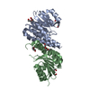
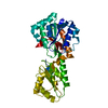
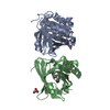
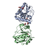
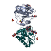
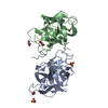
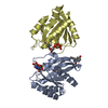
- PDB-5yba: Dimeric Cyclophilin from T.vaginalis in complex with Myb1 peptide -
+ Open data

- Basic information
| Entry | Database: PDB / ID: 5yba | ||||||
|---|---|---|---|---|---|---|---|
| Title | Dimeric Cyclophilin from T.vaginalis in complex with Myb1 peptide | ||||||
 Components Components |
| ||||||
 Keywords Keywords |  ISOMERASE / Divergent loop cyclophilin / Cyclophilin A / Rotamase complex ISOMERASE / Divergent loop cyclophilin / Cyclophilin A / Rotamase complex | ||||||
| Function / homology |  Function and homology informationcyclosporin A binding / protein peptidyl-prolyl isomerization / peptidylprolyl isomerase / peptidyl-prolyl cis-trans isomerase activity / protein folding / DNA-binding transcription factor activity, RNA polymerase II-specific / RNA polymerase II cis-regulatory region sequence-specific DNA binding / regulation of DNA-templated transcription / nucleus / cytoplasm Function and homology informationcyclosporin A binding / protein peptidyl-prolyl isomerization / peptidylprolyl isomerase / peptidyl-prolyl cis-trans isomerase activity / protein folding / DNA-binding transcription factor activity, RNA polymerase II-specific / RNA polymerase II cis-regulatory region sequence-specific DNA binding / regulation of DNA-templated transcription / nucleus / cytoplasmSimilarity search - Function | ||||||
| Biological species |  Trichomonas vaginalis (eukaryote) Trichomonas vaginalis (eukaryote) | ||||||
| Method | X-RAY DIFFRACTION / SYNCHROTRON / MOLECULAR REPLACEMENT / Resolution: 2.062 Å | ||||||
 Authors Authors | Cho, C.C. / Lin, M.H. / Martin, T. / Chou, C.C. / Chen, C. / Hsu, C.H. | ||||||
 Citation Citation | Journal: Sci Rep / Year: 2018 Title: Structural basis of interaction between dimeric cyclophilin 1 and Myb1 transcription factor in Trichomonas vaginalis Authors: Martin, T. / Lou, Y.C. / Chou, C.C. / Wei, S.Y. / Sadotra, S. / Cho, C.C. / Lin, M.H. / Tai, J.H. / Hsu, C.H. / Chen, C. | ||||||
| History |
|
- Structure visualization
| Structure viewer | Molecule: MolmilJmol/JSmol |
|---|
- Downloads & links
-Download
| PDBx/mmCIF format | 5yba.cif.gz | 85.3 KB | Display | PDBx/mmCIF format |
|---|---|---|---|---|
| PDB format | pdb5yba.ent.gz | 63 KB | Display | PDB format |
| PDBx/mmJSON format | 5yba.json.gz | Tree view | PDBx/mmJSON format | |
| Others |  Other downloads Other downloads |
-Validation report
| Arichive directory | https://data.pdbj.org/pub/pdb/validation_reports/yb/5ybaftp://data.pdbj.org/pub/pdb/validation_reports/yb/5yba | HTTPS FTP |
|---|
-Related structure data
| Related structure data |  5yb9C 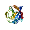 1dywS S: Starting model for refinement C: citing same article ( |
|---|---|
| Similar structure data |


-Links
 PDBj
PDBj

- Assembly
| Deposited unit | 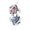
| ||||||||
|---|---|---|---|---|---|---|---|---|---|
| 1 | 
| ||||||||
| Unit cell |
|
-Components
| #1: Protein/peptide | Mass: 1023.142 Da / Num. of mol.: 2 / Source method: obtained synthetically / Details: Minimum Binding Sequence of Myb1 / Source: (synth.) Trichomonas vaginalis (eukaryote) / References: UniProt: Q58HP2*PLUS#2: Protein | Prolyl isomerase / PPIaseMass: 19358.309 Da / Num. of mol.: 2 Source method: isolated from a genetically manipulated source Details: human parasite / Source: (gene. exp.) Trichomonas vaginalis (eukaryote) / Gene: TVAG_004440 / Plasmid: pET28a / Production host:  Escherichia coli BL21(DE3) (bacteria) / Strain (production host): BL21(DE3) / References: UniProt: A2DT06, peptidylprolyl isomerase Escherichia coli BL21(DE3) (bacteria) / Strain (production host): BL21(DE3) / References: UniProt: A2DT06, peptidylprolyl isomerase#3: Water | ChemComp-HOH / | Water Mass: 18.015 Da / Num. of mol.: 161 / Source method: isolated from a natural source / Formula: H2O Mass: 18.015 Da / Num. of mol.: 161 / Source method: isolated from a natural source / Formula: H2O |
|---|
-Experimental details
-Experiment
| Experiment | Method: X-RAY DIFFRACTION / Number of used crystals: 1 |
|---|
- Sample preparation
| Crystal | Density Matthews: 2.13 Å3/Da / Density % sol: 42.24 % |
|---|---|
| Crystal grow | Temperature: 283 K / Method: vapor diffusion, sitting drop / pH: 8 Details: 100 mM Tris-HCl pH 8.0, 30% (v/v) polyethylene glycol 400 |
-Data collection
| Diffraction | Mean temperature: 100 K |
|---|---|
| Diffraction source | Source: SYNCHROTRON / Site: NSRRC  / Beamline: BL13C1 / Wavelength: 0.9762 Å / Beamline: BL13C1 / Wavelength: 0.9762 Å |
| Detector | Type: ADSC QUANTUM 315r / Detector: CCD / Date: Nov 17, 2016 |
| Radiation | Protocol: SINGLE WAVELENGTH / Monochromatic (M) / Laue (L): M / Scattering type: x-ray |
| Radiation wavelength | Wavelength: 0.9762 Å / Relative weight: 1 |
| Reflection | Resolution: 2.06→29.45 Å / Num. obs: 22285 / % possible obs: 91 % / Redundancy: 5.3 % / Net I/σ(I): 9.81 |
| Reflection shell | Resolution: 2.06→2.13 Å / Redundancy: 3.9 % / Rmerge(I) obs: 0.543 / Mean I/σ(I) obs: 2.039 / Rsym value: 0.543 / % possible all: 90.3 |
- Processing
| Software |
| ||||||||||||||||||||||||||||||||||||||||||||||||||||||||||||||||||||||||||||||||||||||||||||||||||||||||||||||||||||||||||||||||||||||||||||||||||||||||||
|---|---|---|---|---|---|---|---|---|---|---|---|---|---|---|---|---|---|---|---|---|---|---|---|---|---|---|---|---|---|---|---|---|---|---|---|---|---|---|---|---|---|---|---|---|---|---|---|---|---|---|---|---|---|---|---|---|---|---|---|---|---|---|---|---|---|---|---|---|---|---|---|---|---|---|---|---|---|---|---|---|---|---|---|---|---|---|---|---|---|---|---|---|---|---|---|---|---|---|---|---|---|---|---|---|---|---|---|---|---|---|---|---|---|---|---|---|---|---|---|---|---|---|---|---|---|---|---|---|---|---|---|---|---|---|---|---|---|---|---|---|---|---|---|---|---|---|---|---|---|---|---|---|---|---|---|
| Refinement | Method to determine structure: MOLECULAR REPLACEMENT Starting model: 1DYW Resolution: 2.062→29.448 Å / SU ML: 0.17 / Cross valid method: FREE R-VALUE / σ(F): 1.34 / Phase error: 22.08
| ||||||||||||||||||||||||||||||||||||||||||||||||||||||||||||||||||||||||||||||||||||||||||||||||||||||||||||||||||||||||||||||||||||||||||||||||||||||||||
| Solvent computation | Shrinkage radii: 0.9 Å / VDW probe radii: 1.11 Å | ||||||||||||||||||||||||||||||||||||||||||||||||||||||||||||||||||||||||||||||||||||||||||||||||||||||||||||||||||||||||||||||||||||||||||||||||||||||||||
| Refinement step | Cycle: LAST / Resolution: 2.062→29.448 Å
| ||||||||||||||||||||||||||||||||||||||||||||||||||||||||||||||||||||||||||||||||||||||||||||||||||||||||||||||||||||||||||||||||||||||||||||||||||||||||||
| Refine LS restraints |
| ||||||||||||||||||||||||||||||||||||||||||||||||||||||||||||||||||||||||||||||||||||||||||||||||||||||||||||||||||||||||||||||||||||||||||||||||||||||||||
| LS refinement shell |
|
