 Movie
Movie Controller

[English] 日本語
 Yorodumi
- PDB-5y7p: Bile salt hydrolase from lactobacillus salivarius complex with gl... -
+ Open data

- Basic information
| Entry | Database: PDB / ID: 5y7p | ||||||
|---|---|---|---|---|---|---|---|
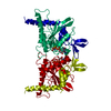
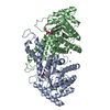
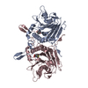
| Title | Bile salt hydrolase from lactobacillus salivarius complex with glycocholic acid and cholic acid | ||||||
 Components Components | Bile salt hydrolase | ||||||
 Keywords Keywords | HYDROLASE / CONJUGATED BILE SALT ACID HYDROLASE / NTN-HYDROLASE / BILE ACIDS / COMPLEX | ||||||
| Function / homology |  Function and homology information Function and homology information | ||||||
| Biological species |  Lactobacillus salivarius (bacteria) Lactobacillus salivarius (bacteria) | ||||||
| Method | X-RAY DIFFRACTION / MOLECULAR REPLACEMENT / Resolution: 2.1 Å | ||||||
 Authors Authors | Hu, X.-J. | ||||||
 Citation Citation | Journal: To Be Published Title: Crystal structure of the bile salt hydrolase from lactobacillus salivarius complex with glycocholic acid and cholic acid Authors: Xu, F. / Hu, X.-J. / Lin, J. | ||||||
| History |
|
- Structure visualization
| Structure viewer | Molecule: MolmilJmol/JSmol |
|---|
- Downloads & links
-Download
| PDBx/mmCIF format | 5y7p.cif.gz | 533.8 KB | Display | PDBx/mmCIF format |
|---|---|---|---|---|
| PDB format | pdb5y7p.ent.gz | 439.8 KB | Display | PDB format |
| PDBx/mmJSON format | 5y7p.json.gz | Tree view | PDBx/mmJSON format | |
| Others |  Other downloads Other downloads |
-Validation report
| Arichive directory | https://data.pdbj.org/pub/pdb/validation_reports/y7/5y7pftp://data.pdbj.org/pub/pdb/validation_reports/y7/5y7p | HTTPS FTP |
|---|
-Related structure data
| Related structure data |  5hkeS S: Starting model for refinement |
|---|---|
| Similar structure data |


-Links
 PDBj
PDBj


- Assembly
| Deposited unit | 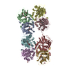
| ||||||||
|---|---|---|---|---|---|---|---|---|---|
| 1 | 
| ||||||||
| 2 | 
| ||||||||
| 3 | 
| ||||||||
| 4 | 
| ||||||||
| Unit cell |
|
-Components
-Protein , 1 types, 8 molecules ABCDEFGH
| #1: Protein | Mass: 37927.285 Da / Num. of mol.: 8 Source method: isolated from a genetically manipulated source Source: (gene. exp.) Lactobacillus salivarius (bacteria) / Production host: Escherichia coli (E. coli) / References: UniProt: J7H3P9 |
|---|
-Non-polymers , 7 types, 1005 molecules 


| #2: Chemical | Glycocholic acid Mass: 465.623 Da / Num. of mol.: 3 / Source method: obtained synthetically / Formula: C26H43NO6 Mass: 465.623 Da / Num. of mol.: 3 / Source method: obtained synthetically / Formula: C26H43NO6#3: Chemical | ChemComp-CHD / Cholic acid Mass: 408.571 Da / Num. of mol.: 6 / Source method: obtained synthetically / Formula: C24H40O5 Mass: 408.571 Da / Num. of mol.: 6 / Source method: obtained synthetically / Formula: C24H40O5#4: Chemical | Glycerol Mass: 92.094 Da / Num. of mol.: 2 / Source method: obtained synthetically / Formula: C3H8O3 Mass: 92.094 Da / Num. of mol.: 2 / Source method: obtained synthetically / Formula: C3H8O3#5: Chemical | ChemComp-PO4 / | Phosphate Mass: 94.971 Da / Num. of mol.: 1 / Source method: obtained synthetically / Formula: PO4 Mass: 94.971 Da / Num. of mol.: 1 / Source method: obtained synthetically / Formula: PO4#6: Chemical | Ethylene glycol Mass: 62.068 Da / Num. of mol.: 3 / Source method: obtained synthetically / Formula: C2H6O2 Mass: 62.068 Da / Num. of mol.: 3 / Source method: obtained synthetically / Formula: C2H6O2#7: Chemical | ChemComp-PEG / | Diethylene glycol Mass: 106.120 Da / Num. of mol.: 1 / Source method: obtained synthetically / Formula: C4H10O3 Mass: 106.120 Da / Num. of mol.: 1 / Source method: obtained synthetically / Formula: C4H10O3#8: Water | ChemComp-HOH / | WaterMass: 18.015 Da / Num. of mol.: 989 / Source method: isolated from a natural source / Formula: H2O |
|---|
-Experimental details
-Experiment
| Experiment | Method: X-RAY DIFFRACTION / Number of used crystals: 1 |
|---|
- Sample preparation
| Crystal | Density Matthews: 2.26 Å3/Da / Density % sol: 45.5 % / Description: shaft |
|---|---|
| Crystal grow | Temperature: 293 K / Method: vapor diffusion, sitting drop / pH: 4.8 Details: 20% polyethylene glycol 3350, 0.2 M potassium dihydrogen phosphate pH 4.8. Then crystal is soaked in this buffer containing 5 uM glycocholic acid for 4 hrs. |
-Data collection
| Diffraction | Mean temperature: 100 K |
|---|---|
| Diffraction source | Source: ROTATING ANODE / Type: RIGAKU FR-E+ DW / Wavelength: 1.5418 Å |
| Detector | Type: RIGAKU SATURN 944 / Detector: CCD / Date: Jul 27, 2015 |
| Radiation | Protocol: SINGLE WAVELENGTH / Monochromatic (M) / Laue (L): M / Scattering type: x-ray |
| Radiation wavelength | Wavelength: 1.5418 Å / Relative weight: 1 |
| Reflection | Resolution: 2.1→50 Å / Num. obs: 143414 / % possible obs: 95.1 % / Redundancy: 5.3 % / Biso Wilson estimate: 18.4 Å2 / Rmerge(I) obs: 0.073 / Net I/σ(I): 12.7 |
| Reflection shell | Resolution: 2.1→2.14 Å / Redundancy: 4.9 % / Num. unique obs: 6716 / % possible all: 89.7 |
- Processing
| Software |
| ||||||||||||||||||||||||||||||||||||||||||||||||||||||||||||||||||||||||||||||||||||||||||||||||||||||||||||||||||||||||||||||||||||||||||||||||||||||||||||||||||||||||||||||||||||||
|---|---|---|---|---|---|---|---|---|---|---|---|---|---|---|---|---|---|---|---|---|---|---|---|---|---|---|---|---|---|---|---|---|---|---|---|---|---|---|---|---|---|---|---|---|---|---|---|---|---|---|---|---|---|---|---|---|---|---|---|---|---|---|---|---|---|---|---|---|---|---|---|---|---|---|---|---|---|---|---|---|---|---|---|---|---|---|---|---|---|---|---|---|---|---|---|---|---|---|---|---|---|---|---|---|---|---|---|---|---|---|---|---|---|---|---|---|---|---|---|---|---|---|---|---|---|---|---|---|---|---|---|---|---|---|---|---|---|---|---|---|---|---|---|---|---|---|---|---|---|---|---|---|---|---|---|---|---|---|---|---|---|---|---|---|---|---|---|---|---|---|---|---|---|---|---|---|---|---|---|---|---|---|---|
| Refinement | Method to determine structure: MOLECULAR REPLACEMENT Starting model: 5HKE Resolution: 2.1→28.5 Å / Cor.coef. Fo:Fc: 0.962 / Cor.coef. Fo:Fc free: 0.934 / SU B: 4.575 / SU ML: 0.121 / Cross valid method: THROUGHOUT / ESU R: 0.236 / ESU R Free: 0.187 / Stereochemistry target values: MAXIMUM LIKELIHOOD / Details: HYDROGENS HAVE BEEN ADDED IN THE RIDING POSITIONS
| ||||||||||||||||||||||||||||||||||||||||||||||||||||||||||||||||||||||||||||||||||||||||||||||||||||||||||||||||||||||||||||||||||||||||||||||||||||||||||||||||||||||||||||||||||||||
| Solvent computation | Ion probe radii: 0.8 Å / Shrinkage radii: 0.8 Å / VDW probe radii: 1.2 Å / Solvent model: MASK | ||||||||||||||||||||||||||||||||||||||||||||||||||||||||||||||||||||||||||||||||||||||||||||||||||||||||||||||||||||||||||||||||||||||||||||||||||||||||||||||||||||||||||||||||||||||
| Displacement parameters | Biso mean: 27.73 Å2
| ||||||||||||||||||||||||||||||||||||||||||||||||||||||||||||||||||||||||||||||||||||||||||||||||||||||||||||||||||||||||||||||||||||||||||||||||||||||||||||||||||||||||||||||||||||||
| Refinement step | Cycle: 1 / Resolution: 2.1→28.5 Å
| ||||||||||||||||||||||||||||||||||||||||||||||||||||||||||||||||||||||||||||||||||||||||||||||||||||||||||||||||||||||||||||||||||||||||||||||||||||||||||||||||||||||||||||||||||||||
| Refine LS restraints |
|