 Movie
Movie Controller


+ Open data

- Basic information
| Entry | Database: PDB / ID: 5y30 | ||||||
|---|---|---|---|---|---|---|---|
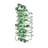
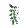
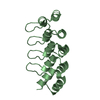
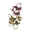
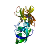
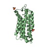
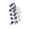
| Title | Crystal structure of LGI1 LRR domain | ||||||
 Components Components | Leucine-rich glioma-inactivated protein 1 | ||||||
 Keywords Keywords |  CELL ADHESION / epilepsy / synapse / ADAM / EPTP / WD40 CELL ADHESION / epilepsy / synapse / ADAM / EPTP / WD40 | ||||||
| Function / homology |  Function and homology information Function and homology informationLGI-ADAM interactions / axon initial segment / neurotransmitter receptor localization to postsynaptic specialization membrane / positive regulation of synaptic transmission / synaptic cleft / myelination / axon guidance / neuron projection development / nervous system development / positive regulation of cell growth ...LGI-ADAM interactions / axon initial segment / neurotransmitter receptor localization to postsynaptic specialization membrane / positive regulation of synaptic transmission / synaptic cleft / myelination / axon guidance / neuron projection development / nervous system development / positive regulation of cell growth / signaling receptor binding / glutamatergic synapse / dendrite / Golgi apparatus / endoplasmic reticulum / extracellular space / extracellular region / cytoplasmSimilarity search - Function | ||||||
| Biological species |  Homo sapiens (human) Homo sapiens (human) | ||||||
| Method | X-RAY DIFFRACTION / SYNCHROTRON / MOLECULAR REPLACEMENT / Resolution: 1.781 Å | ||||||
 Authors Authors | Yamagata, A. / Fukai, S. | ||||||
 Citation Citation | Journal: Nat Commun / Year: 2018 Title: Structural basis of epilepsy-related ligand-receptor complex LGI1-ADAM22. Authors: Yamagata, A. / Miyazaki, Y. / Yokoi, N. / Shigematsu, H. / Sato, Y. / Goto-Ito, S. / Maeda, A. / Goto, T. / Sanbo, M. / Hirabayashi, M. / Shirouzu, M. / Fukata, Y. / Fukata, M. / Fukai, S. | ||||||
| History |
|
- Structure visualization
| Structure viewer | Molecule: MolmilJmol/JSmol |
|---|
- Downloads & links
-Download
| PDBx/mmCIF format | 5y30.cif.gz | 93.2 KB | Display | PDBx/mmCIF format |
|---|---|---|---|---|
| PDB format | pdb5y30.ent.gz | 69.3 KB | Display | PDB format |
| PDBx/mmJSON format | 5y30.json.gz | Tree view | PDBx/mmJSON format | |
| Others |  Other downloads Other downloads |
-Validation report
| Arichive directory | https://data.pdbj.org/pub/pdb/validation_reports/y3/5y30ftp://data.pdbj.org/pub/pdb/validation_reports/y3/5y30 | HTTPS FTP |
|---|
-Related structure data
| Related structure data |  5y2zC 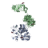 5y31C 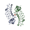 2wfhS S: Starting model for refinement C: citing same article ( |
|---|---|
| Similar structure data |


-Links
 PDBj
PDBj


- Assembly
| Deposited unit | 
| ||||||||
|---|---|---|---|---|---|---|---|---|---|
| 1 |
| ||||||||
| Unit cell |
|
-Components
| #1: Protein | Mass: 23808.107 Da / Num. of mol.: 1 / Fragment: LGI1 LRR domain (UNP RESIDUES 37-223) Source method: isolated from a genetically manipulated source Source: (gene. exp.) Homo sapiens (human) / Gene: LGI1, EPT, UNQ775/PRO1569 / Cell line (production host): Expi293F / Production host: Homo sapiens (human) / References: UniProt: O95970 |
|---|---|
| #2: Water | ChemComp-HOH / Water Mass: 18.015 Da / Num. of mol.: 100 / Source method: isolated from a natural source / Formula: H2O Mass: 18.015 Da / Num. of mol.: 100 / Source method: isolated from a natural source / Formula: H2O |
-Experimental details
-Experiment
| Experiment | Method: X-RAY DIFFRACTION / Number of used crystals: 1 |
|---|
- Sample preparation
| Crystal | Density Matthews: 2.83 Å3/Da / Density % sol: 56.58 % |
|---|---|
| Crystal grow | Temperature: 293 K / Method: vapor diffusion, sitting drop / pH: 5.5 Details: 20% PEG 4000, 0.2 M ammonium acetate, 0.1 M tri-sodium citrate (pH 5.5) |
-Data collection
| Diffraction | Mean temperature: 100 K |
|---|---|
| Diffraction source | Source: SYNCHROTRON / Site: SPring-8  / Beamline: BL41XU / Wavelength: 1 Å / Beamline: BL41XU / Wavelength: 1 Å |
| Detector | Type: DECTRIS PILATUS3 S 6M / Detector: PIXEL / Date: Oct 7, 2015 |
| Radiation | Protocol: SINGLE WAVELENGTH / Monochromatic (M) / Laue (L): M / Scattering type: x-ray |
| Radiation wavelength | Wavelength: 1 Å / Relative weight: 1 |
| Reflection | Resolution: 1.78→50 Å / Num. obs: 25330 / % possible obs: 99.9 % / Redundancy: 15.1 % / Rsym value: 0.055 / Net I/σ(I): 47.2 |
| Reflection shell | Resolution: 1.78→1.81 Å / Mean I/σ(I) obs: 2.2 / CC1/2: 0.558 / Rsym value: 0.45 / % possible all: 99.8 |
- Processing
| Software |
| ||||||||||||||||||||||||||||||||||||||||||||||||||||||||||||||||||||||
|---|---|---|---|---|---|---|---|---|---|---|---|---|---|---|---|---|---|---|---|---|---|---|---|---|---|---|---|---|---|---|---|---|---|---|---|---|---|---|---|---|---|---|---|---|---|---|---|---|---|---|---|---|---|---|---|---|---|---|---|---|---|---|---|---|---|---|---|---|---|---|---|
| Refinement | Method to determine structure: MOLECULAR REPLACEMENT Starting model: 2WFH Resolution: 1.781→39.366 Å / SU ML: 0.2 / Cross valid method: FREE R-VALUE / σ(F): 1.49 / Phase error: 20.38
| ||||||||||||||||||||||||||||||||||||||||||||||||||||||||||||||||||||||
| Solvent computation | Shrinkage radii: 0.9 Å / VDW probe radii: 1.11 Å | ||||||||||||||||||||||||||||||||||||||||||||||||||||||||||||||||||||||
| Refinement step | Cycle: LAST / Resolution: 1.781→39.366 Å
| ||||||||||||||||||||||||||||||||||||||||||||||||||||||||||||||||||||||
| Refine LS restraints |
| ||||||||||||||||||||||||||||||||||||||||||||||||||||||||||||||||||||||
| LS refinement shell |
| ||||||||||||||||||||||||||||||||||||||||||||||||||||||||||||||||||||||
| Refinement TLS params. | Method: refined / Origin x: -1.5236 Å / Origin y: 21.0476 Å / Origin z: -0.7258 Å
| ||||||||||||||||||||||||||||||||||||||||||||||||||||||||||||||||||||||
| Refinement TLS group | Selection details: all |
