 Movie
Movie Controller


+ Open data

- Basic information
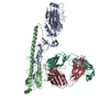
| Entry | Database: PDB / ID: 5xwd | |||||||||
|---|---|---|---|---|---|---|---|---|---|---|
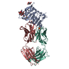
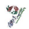
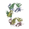
| Title | Crystal structure of the complex of 059-152-Fv and EGFR-ECD | |||||||||
 Components Components |
| |||||||||
 Keywords Keywords |  SIGNALING PROTEIN / Antibody / Receptor / Complex SIGNALING PROTEIN / Antibody / Receptor / Complex | |||||||||
| Function / homology |  Function and homology information Function and homology informationresponse to hydroxyisoflavone / multivesicular body, internal vesicle lumen / positive regulation of prolactin secretion / negative regulation of cardiocyte differentiation / positive regulation of protein kinase C activity / diterpenoid metabolic process / Shc-EGFR complex / ovulation cycle / Inhibition of Signaling by Overexpressed EGFR / epidermal growth factor receptor activity ...response to hydroxyisoflavone / multivesicular body, internal vesicle lumen / positive regulation of prolactin secretion / negative regulation of cardiocyte differentiation / positive regulation of protein kinase C activity / diterpenoid metabolic process / Shc-EGFR complex / ovulation cycle / Inhibition of Signaling by Overexpressed EGFR / epidermal growth factor receptor activity / EGFR interacts with phospholipase C-gamma / positive regulation of mucus secretion / response to UV-A / epidermal growth factor binding / PLCG1 events in ERBB2 signaling / tongue development / midgut development / ERBB2-EGFR signaling pathway / hydrogen peroxide metabolic process / PTK6 promotes HIF1A stabilization / digestive tract morphogenesis / regulation of nitric-oxide synthase activity / morphogenesis of an epithelial fold / ERBB2 Activates PTK6 Signaling / intracellular vesicle / Signaling by EGFR / response to cobalamin / transmembrane receptor protein tyrosine kinase activator activity / protein tyrosine kinase activator activity / negative regulation of epidermal growth factor receptor signaling pathway / Signaling by ERBB4 / regulation of phosphatidylinositol 3-kinase/protein kinase B signal transduction / eyelid development in camera-type eye / protein insertion into membrane / cerebral cortex cell migration / ERBB2 Regulates Cell Motility / regulation of JNK cascade / : / PI3K events in ERBB2 signaling / positive regulation of cyclin-dependent protein serine/threonine kinase activity / negative regulation of mitotic cell cycle / hair follicle development / MAP kinase kinase kinase activity / Estrogen-dependent nuclear events downstream of ESR-membrane signaling / embryonic placenta development / positive regulation of bone resorption / positive regulation of G1/S transition of mitotic cell cycle / GAB1 signalosome / positive regulation of nitric oxide mediated signal transduction / salivary gland morphogenesis / peptidyl-tyrosine autophosphorylation / regulation of peptidyl-tyrosine phosphorylation / positive regulation of phosphorylation / positive regulation of glial cell proliferation / positive regulation of vasoconstriction / Signaling by ERBB2 / cellular response to epidermal growth factor stimulus / cellular response to cadmium ion / GRB2 events in EGFR signaling / SHC1 events in EGFR signaling / positive regulation of DNA repair / EGFR Transactivation by Gastrin / GRB2 events in ERBB2 signaling / TFAP2 (AP-2) family regulates transcription of growth factors and their receptors / transmembrane receptor protein tyrosine kinase activity / SHC1 events in ERBB2 signaling / ossification / positive regulation of synaptic transmission, glutamatergic / neurogenesis / cellular response to dexamethasone stimulus / basal plasma membrane / regulation of ERK1 and ERK2 cascade / neuron projection morphogenesis / positive regulation of superoxide anion generation / positive regulation of DNA replication / Signal transduction by L1 / epithelial cell proliferation / cellular response to estradiol stimulus / NOTCH3 Activation and Transmission of Signal to the Nucleus / positive regulation of epithelial cell proliferation / astrocyte activation / liver regeneration / positive regulation of protein localization to plasma membrane / EGFR downregulation / cell surface receptor protein tyrosine kinase signaling pathway / cellular response to amino acid stimulus / positive regulation of smooth muscle cell proliferation / Signaling by ERBB2 TMD/JMD mutants / positive regulation of MAP kinase activity / clathrin-coated endocytic vesicle membrane / lung development / Constitutive Signaling by EGFRvIII / Signaling by ERBB2 ECD mutants / epidermal growth factor receptor signaling pathway / Signaling by ERBB2 KD Mutants / negative regulation of protein catabolic process / receptor protein-tyrosine kinase / Downregulation of ERBB2 signaling / kinase binding / ruffle membraneSimilarity search - Function | |||||||||
| Biological species |  Homo sapiens (human) Homo sapiens (human) | |||||||||
| Method | X-RAY DIFFRACTION / SYNCHROTRON / MOLECULAR REPLACEMENT / Resolution: 2.894 Å | |||||||||
 Authors Authors | Matsuda, T. / Ito, T. / Shirouzu, M. | |||||||||
 Citation Citation | Journal: PLoS ONE / Year: 2018 Title: Cell-free synthesis of functional antibody fragments to provide a structural basis for antibody-antigen interaction Authors: Matsuda, T. / Ito, T. / Takemoto, C. / Katsura, K. / Ikeda, M. / Wakiyama, M. / Kukimoto-Niino, M. / Yokoyama, S. / Kurosawa, Y. / Shirouzu, M. | |||||||||
| History |
|
- Structure visualization
| Structure viewer | Molecule: MolmilJmol/JSmol |
|---|
- Downloads & links
-Download
| PDBx/mmCIF format | 5xwd.cif.gz | 184.1 KB | Display | PDBx/mmCIF format |
|---|---|---|---|---|
| PDB format | pdb5xwd.ent.gz | 141.3 KB | Display | PDB format |
| PDBx/mmJSON format | 5xwd.json.gz | Tree view | PDBx/mmJSON format | |
| Others |  Other downloads Other downloads |
-Validation report
| Arichive directory | https://data.pdbj.org/pub/pdb/validation_reports/xw/5xwdftp://data.pdbj.org/pub/pdb/validation_reports/xw/5xwd | HTTPS FTP |
|---|
-Related structure data


-Links
 PDBj
PDBj


- Assembly
| Deposited unit | 
| ||||||||
|---|---|---|---|---|---|---|---|---|---|
| 1 |
| ||||||||
| Unit cell |
|
-Components
-Antibody , 2 types, 2 molecules HD
| #2: Antibody | Mass: 14017.562 Da / Num. of mol.: 1 Source method: isolated from a genetically manipulated source Source: (gene. exp.) Homo sapiens (human) / Description: cell-free synthesis based on E.coli extract / Production host: cell-free synthesis (others) |
|---|---|
| #3: Antibody | Mass: 12509.670 Da / Num. of mol.: 1 Source method: isolated from a genetically manipulated source Source: (gene. exp.) Homo sapiens (human) / Description: cell-free synthesis based on E.coli extract / Production host: cell-free synthesis (others) |
-Protein / Non-polymers , 2 types, 21 molecules A
| #1: Protein | / Proto-oncogene c-ErbB-1 / Receptor tyrosine-protein kinase erbB-1 Mass: 71688.695 Da / Num. of mol.: 1 / Fragment: UNP residues 1-643 Source method: isolated from a genetically manipulated source Source: (gene. exp.) Homo sapiens (human) / Gene: EGFR, ERBB, ERBB1, HER1 / Production host: Homo sapiens (human)References: UniProt: P00533, receptor protein-tyrosine kinase |
|---|---|
| #6: Chemical | ChemComp-ZN /  Mass: 65.409 Da / Num. of mol.: 20 Mass: 65.409 Da / Num. of mol.: 20Source method: isolated from a genetically manipulated source Formula: Zn |
-Sugars , 2 types, 8 molecules 
| #4: Polysaccharide | 2-acetamido-2-deoxy-beta-D-glucopyranose-(1-2)-alpha-D-mannopyranose-(1-3)-[2-acetamido-2-deoxy- ...2-acetamido-2-deoxy-beta-D-glucopyranose-(1-2)-alpha-D-mannopyranose-(1-3)-[2-acetamido-2-deoxy-beta-D-glucopyranose-(1-2)-alpha-D-mannopyranose-(1-6)]beta-D-mannopyranose-(1-4)-2-acetamido-2-deoxy-beta-D-glucopyranose-(1-4)-2-acetamido-2-deoxy-beta-D-glucopyranose / Mass: 1317.209 Da / Num. of mol.: 1 / Source method: obtained synthetically |
|---|---|
| #5: Sugar | ChemComp-NAG / N-Acetylglucosamine Type: D-saccharide, beta linking / Mass: 221.208 Da / Num. of mol.: 7 / Source method: obtained synthetically / Formula: C8H15NO6 Type: D-saccharide, beta linking / Mass: 221.208 Da / Num. of mol.: 7 / Source method: obtained synthetically / Formula: C8H15NO6 |
-Experimental details
-Experiment
| Experiment | Method: X-RAY DIFFRACTION / Number of used crystals: 1 |
|---|
- Sample preparation
| Crystal | Density Matthews: 3.54 Å3/Da / Density % sol: 65.23 % |
|---|---|
| Crystal grow | Temperature: 293 K / Method: vapor diffusion, sitting drop / pH: 6 / Details: MES buffer (pH 6.0), zinc acetate, PEG8000 |
-Data collection
| Diffraction | Mean temperature: 100 K |
|---|---|
| Diffraction source | Source: SYNCHROTRON / Site: Photon Factory  / Beamline: AR-NE3A / Wavelength: 1 Å / Beamline: AR-NE3A / Wavelength: 1 Å |
| Detector | Type: ADSC QUANTUM 270 / Detector: CCD / Date: Feb 16, 2014 |
| Radiation | Protocol: SINGLE WAVELENGTH / Monochromatic (M) / Laue (L): M / Scattering type: x-ray |
| Radiation wavelength | Wavelength: 1 Å / Relative weight: 1 |
| Reflection | Resolution: 2.894→50 Å / Num. obs: 680489 / % possible obs: 99.6 % / Redundancy: 21.7 % / Net I/σ(I): 39.8 |
| Reflection shell | Resolution: 2.9→3 Å / Redundancy: 22.1 % / Rsym value: 2.053 / % possible all: 100 |
- Processing
| Software |
| ||||||||||||||||||||||||||||||||||||||||||||||||||||||||||||||||||||||||||||||||||||
|---|---|---|---|---|---|---|---|---|---|---|---|---|---|---|---|---|---|---|---|---|---|---|---|---|---|---|---|---|---|---|---|---|---|---|---|---|---|---|---|---|---|---|---|---|---|---|---|---|---|---|---|---|---|---|---|---|---|---|---|---|---|---|---|---|---|---|---|---|---|---|---|---|---|---|---|---|---|---|---|---|---|---|---|---|---|
| Refinement | Method to determine structure: MOLECULAR REPLACEMENT Starting model: 3NJP, 3B2U Resolution: 2.894→32.494 Å / SU ML: 0.58 / Cross valid method: FREE R-VALUE / σ(F): 1.33 / Phase error: 38.83
| ||||||||||||||||||||||||||||||||||||||||||||||||||||||||||||||||||||||||||||||||||||
| Solvent computation | Shrinkage radii: 0.9 Å / VDW probe radii: 1.11 Å | ||||||||||||||||||||||||||||||||||||||||||||||||||||||||||||||||||||||||||||||||||||
| Refinement step | Cycle: LAST / Resolution: 2.894→32.494 Å
| ||||||||||||||||||||||||||||||||||||||||||||||||||||||||||||||||||||||||||||||||||||
| Refine LS restraints |
| ||||||||||||||||||||||||||||||||||||||||||||||||||||||||||||||||||||||||||||||||||||
| LS refinement shell |
|
