 Movie
Movie Controller

[English] 日本語
 Yorodumi
- PDB-5xj5: Crystal structure of PlsY (YgiH), an integral membrane glycerol 3... -
+ Open data

- Basic information
| Entry | Database: PDB / ID: 5xj5 | |||||||||||||||||||||
|---|---|---|---|---|---|---|---|---|---|---|---|---|---|---|---|---|---|---|---|---|---|---|
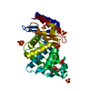
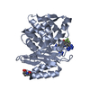
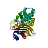
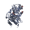
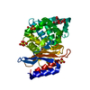
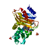
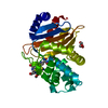
| Title | Crystal structure of PlsY (YgiH), an integral membrane glycerol 3-phosphate acyltransferase - the monoacylglycerol form | |||||||||||||||||||||
 Components Components | Glycerol-3-phosphate acyltransferase | |||||||||||||||||||||
 Keywords Keywords |  TRANSFERASE / glycerol 3-phosphate acyltransferase / glycerylphosphate acyltransferase / GPAT / in meso / lipid cubic phase / lipidic cubic phase / lipid metabolism / monoacylglycerol / phospholipid biosynthesis / PlsY / YgiH TRANSFERASE / glycerol 3-phosphate acyltransferase / glycerylphosphate acyltransferase / GPAT / in meso / lipid cubic phase / lipidic cubic phase / lipid metabolism / monoacylglycerol / phospholipid biosynthesis / PlsY / YgiH | |||||||||||||||||||||
| Function / homology |  Function and homology information Function and homology informationacyl phosphate:glycerol-3-phosphate acyltransferase / acyl-phosphate glycerol-3-phosphate acyltransferase activity / phospholipid biosynthetic process / plasma membraneSimilarity search - Function | |||||||||||||||||||||
| Biological species |   Aquifex aeolicus (bacteria) Aquifex aeolicus (bacteria) | |||||||||||||||||||||
| Method | X-RAY DIFFRACTION / SYNCHROTRON / SAD / Resolution: 1.481 Å | |||||||||||||||||||||
 Authors Authors | Li, Z. / Li, D. | |||||||||||||||||||||
| Funding support |  China, 6items China, 6items
| |||||||||||||||||||||
 Citation Citation | Journal: Nat Commun / Year: 2017 Title: Structural insights into the committed step of bacterial phospholipid biosynthesis. Authors: Li, Z. / Tang, Y. / Wu, Y. / Zhao, S. / Bao, J. / Luo, Y. / Li, D. | |||||||||||||||||||||
| History |
|
- Structure visualization
| Structure viewer | Molecule: MolmilJmol/JSmol |
|---|
- Downloads & links
-Download
| PDBx/mmCIF format | 5xj5.cif.gz | 59.1 KB | Display | PDBx/mmCIF format |
|---|---|---|---|---|
| PDB format | pdb5xj5.ent.gz | 48.2 KB | Display | PDB format |
| PDBx/mmJSON format | 5xj5.json.gz | Tree view | PDBx/mmJSON format | |
| Others |  Other downloads Other downloads |
-Validation report
| Arichive directory | https://data.pdbj.org/pub/pdb/validation_reports/xj/5xj5ftp://data.pdbj.org/pub/pdb/validation_reports/xj/5xj5 | HTTPS FTP |
|---|
-Related structure data


-Links
 PDBj
PDBj

- Assembly
| Deposited unit | 
| ||||||||
|---|---|---|---|---|---|---|---|---|---|
| 1 |
| ||||||||
| Unit cell |
|
-Components
-Protein , 1 types, 1 molecules A
| #1: Protein | Mass: 21892.002 Da / Num. of mol.: 1 Source method: isolated from a genetically manipulated source Source: (gene. exp.) Aquifex aeolicus (bacteria) / Strain: VF5 / Gene: plsY, aq_676 / Plasmid: p3EGDetails (production host): pET backbone with eGFP fusion at C terminal Production host: Escherichia coli (E. coli) / Strain (production host): BL21(DE3)References: UniProt: O66905, Transferases; Acyltransferases; Transferring groups other than aminoacyl groups |
|---|
-Non-polymers , 5 types, 97 molecules 


| #2: Chemical | ChemComp-SO4 / Sulfate Mass: 96.063 Da / Num. of mol.: 7 / Source method: obtained synthetically / Formula: SO4 Mass: 96.063 Da / Num. of mol.: 7 / Source method: obtained synthetically / Formula: SO4#3: Chemical | ChemComp-78M / (  Mass: 314.460 Da / Num. of mol.: 15 / Source method: obtained synthetically / Formula: C18H34O4 Mass: 314.460 Da / Num. of mol.: 15 / Source method: obtained synthetically / Formula: C18H34O4#4: Chemical | ChemComp-PGE / | Polyethylene glycol Mass: 150.173 Da / Num. of mol.: 1 / Source method: obtained synthetically / Formula: C6H14O4 Mass: 150.173 Da / Num. of mol.: 1 / Source method: obtained synthetically / Formula: C6H14O4#5: Chemical | ChemComp-GLY / | Glycine Type: peptide linking / Mass: 75.067 Da / Num. of mol.: 1 / Source method: obtained synthetically / Formula: C2H5NO2 Type: peptide linking / Mass: 75.067 Da / Num. of mol.: 1 / Source method: obtained synthetically / Formula: C2H5NO2#6: Water | ChemComp-HOH / | WaterMass: 18.015 Da / Num. of mol.: 73 / Source method: isolated from a natural source / Formula: H2O |
|---|
-Experimental details
-Experiment
| Experiment | Method: X-RAY DIFFRACTION / Number of used crystals: 1 |
|---|
- Sample preparation
| Crystal | Density Matthews: 2.96 Å3/Da / Density % sol: 58.39 % |
|---|---|
| Crystal grow | Temperature: 293 K / Method: lipidic cubic phase / pH: 3.8 Details: 7.8 monoacylglycerol (7.8 MAG), 0.1 M ammonium sulphate, 25-30% triethylene glycole, 0.1 M glycine/HCl pH 3.8 |
-Data collection
| Diffraction | Mean temperature: 100 K |
|---|---|
| Diffraction source | Source: SYNCHROTRON / Site: NFPSS / Beamline: BL19U1 / Wavelength: 0.97852 Å |
| Detector | Type: DECTRIS PILATUS3 6M / Detector: PIXEL / Date: Sep 9, 2016 |
| Radiation | Protocol: SINGLE WAVELENGTH / Monochromatic (M) / Laue (L): M / Scattering type: x-ray |
| Radiation wavelength | Wavelength: 0.97852 Å / Relative weight: 1 |
| Reflection | Resolution: 1.48→47.44 Å / Num. obs: 43520 / % possible obs: 98.4 % / Redundancy: 5.6 % / Rmerge(I) obs: 0.054 / Rpim(I) all: 0.025 / Net I/σ(I): 16.6 |
| Reflection shell | Resolution: 1.48→1.51 Å / Redundancy: 5.1 % / Rmerge(I) obs: 0.893 / Mean I/σ(I) obs: 1.7 / Rpim(I) all: 0.428 / % possible all: 94.4 |
- Processing
| Software |
| ||||||||||||||||||||||||||||||||||||||||||||||||||||||||||||||||||||||||||||||||||||||||||||||||||||||||||||||||
|---|---|---|---|---|---|---|---|---|---|---|---|---|---|---|---|---|---|---|---|---|---|---|---|---|---|---|---|---|---|---|---|---|---|---|---|---|---|---|---|---|---|---|---|---|---|---|---|---|---|---|---|---|---|---|---|---|---|---|---|---|---|---|---|---|---|---|---|---|---|---|---|---|---|---|---|---|---|---|---|---|---|---|---|---|---|---|---|---|---|---|---|---|---|---|---|---|---|---|---|---|---|---|---|---|---|---|---|---|---|---|---|---|---|
| Refinement | Method to determine structure: SAD / Resolution: 1.481→35.582 Å / SU ML: 0.15 / Cross valid method: FREE R-VALUE / σ(F): 1.34 / Phase error: 22.13
| ||||||||||||||||||||||||||||||||||||||||||||||||||||||||||||||||||||||||||||||||||||||||||||||||||||||||||||||||
| Solvent computation | Shrinkage radii: 0.9 Å / VDW probe radii: 1.11 Å | ||||||||||||||||||||||||||||||||||||||||||||||||||||||||||||||||||||||||||||||||||||||||||||||||||||||||||||||||
| Displacement parameters | Biso mean: 27.53 Å2 | ||||||||||||||||||||||||||||||||||||||||||||||||||||||||||||||||||||||||||||||||||||||||||||||||||||||||||||||||
| Refinement step | Cycle: LAST / Resolution: 1.481→35.582 Å
| ||||||||||||||||||||||||||||||||||||||||||||||||||||||||||||||||||||||||||||||||||||||||||||||||||||||||||||||||
| Refine LS restraints |
| ||||||||||||||||||||||||||||||||||||||||||||||||||||||||||||||||||||||||||||||||||||||||||||||||||||||||||||||||
| LS refinement shell |
|
