 Movie
Movie Controller


+ Open data

- Basic information
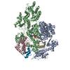
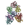
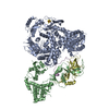
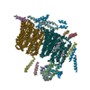
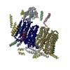
| Entry | Database: PDB / ID: 5xeq | |||||||||
|---|---|---|---|---|---|---|---|---|---|---|
| Title | Crystal Structure of human MDGA1 and human neuroligin-2 complex | |||||||||
 Components Components |
| |||||||||
 Keywords Keywords |  CELL ADHESION / Immunogloubulin-like domain CELL ADHESION / Immunogloubulin-like domain | |||||||||
| Function / homology |  Function and homology information Function and homology informationneurotransmitter-gated ion channel clustering / jump response / positive regulation of t-SNARE clustering / symmetric, GABA-ergic, inhibitory synapse / cerebral cortex radially oriented cell migration / spinal cord association neuron differentiation / gephyrin clustering involved in postsynaptic density assembly / terminal button organization / postsynaptic density protein 95 clustering / postsynaptic membrane assembly ...neurotransmitter-gated ion channel clustering / jump response / positive regulation of t-SNARE clustering / symmetric, GABA-ergic, inhibitory synapse / cerebral cortex radially oriented cell migration / spinal cord association neuron differentiation / gephyrin clustering involved in postsynaptic density assembly / terminal button organization / postsynaptic density protein 95 clustering / postsynaptic membrane assembly / cell-cell junction maintenance / positive regulation of synaptic vesicle clustering / presynaptic membrane assembly / positive regulation of inhibitory postsynaptic potential / thigmotaxis / ribbon synapse / Post-translational modification: synthesis of GPI-anchored proteins / inhibitory synapse / neuron cell-cell adhesion / insulin metabolic process / regulation of respiratory gaseous exchange by nervous system process / neurexin family protein binding / dopaminergic synapse / presynapse assembly / inhibitory synapse assembly / protein localization to synapse / glycinergic synapse / regulation of AMPA receptor activity / postsynaptic specialization membrane / synaptic transmission, GABAergic / protein localization to cell surface / Neurexins and neuroligins / positive regulation of synapse assembly / positive regulation of protein localization to synapse / regulation of synaptic membrane adhesion / positive regulation of dendritic spine development / locomotory exploration behavior / social behavior / positive regulation of excitatory postsynaptic potential / neuromuscular process controlling balance / regulation of presynapse assembly / excitatory synapse / synaptic vesicle endocytosis / GABA-ergic synapse / side of membrane / sensory perception of pain / synapse assembly / cell adhesion molecule binding / positive regulation of synaptic transmission, glutamatergic / dendritic shaft / positive regulation of synaptic transmission, GABAergic / synapse organization / brain development / neuron migration / modulation of chemical synaptic transmission / positive regulation of insulin secretion / cell-cell adhesion / signaling receptor activity / presynaptic membrane / nervous system development / chemical synaptic transmission / postsynaptic membrane / synapse / positive regulation of cell population proliferation / Golgi apparatus / cell surface / extracellular space / extracellular region / membrane / identical protein binding / plasma membraneSimilarity search - Function | |||||||||
| Biological species |  Homo sapiens (human) Homo sapiens (human) | |||||||||
| Method | X-RAY DIFFRACTION / SYNCHROTRON / MOLECULAR REPLACEMENT / Resolution: 3.136 Å | |||||||||
 Authors Authors | Kim, H.M. / Kim, J.A. / Kim, D. | |||||||||
 Citation Citation | Journal: Neuron / Year: 2017 Title: Structural Insights into Modulation of Neurexin-Neuroligin Trans-synaptic Adhesion by MDGA1/Neuroligin-2 Complex Authors: Kim, J.A. / Kim, D. / Won, S.Y. / Han, K.A. / Park, D. / Cho, E. / Yun, N. / An, H.J. / Um, J.W. / Kim, E. / Lee, J.O. / Ko, J. / Kim, H.M. | |||||||||
| History |
|
- Structure visualization
| Structure viewer | Molecule: MolmilJmol/JSmol |
|---|
- Downloads & links
-Download
| PDBx/mmCIF format | 5xeq.cif.gz | 176.9 KB | Display | PDBx/mmCIF format |
|---|---|---|---|---|
| PDB format | pdb5xeq.ent.gz | 136 KB | Display | PDB format |
| PDBx/mmJSON format | 5xeq.json.gz | Tree view | PDBx/mmJSON format | |
| Others |  Other downloads Other downloads |
-Validation report
| Arichive directory | https://data.pdbj.org/pub/pdb/validation_reports/xe/5xeqftp://data.pdbj.org/pub/pdb/validation_reports/xe/5xeq | HTTPS FTP |
|---|
-Related structure data
| Related structure data |  3bl8S S: Starting model for refinement |
|---|---|
| Similar structure data |


-Links
 PDBj
PDBj


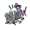
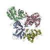
- Assembly
| Deposited unit | 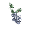
| ||||||||
|---|---|---|---|---|---|---|---|---|---|
| 1 | 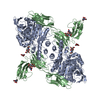
| ||||||||
| Unit cell |
|
-Components
| #1: Protein | Mass: 64101.059 Da / Num. of mol.: 1 / Fragment: UNP residues 42-610 / Mutation: N98Q Source method: isolated from a genetically manipulated source Source: (gene. exp.) Homo sapiens (human) / Gene: NLGN2, KIAA1366 / Plasmid: pAcGP67A / Production host:   Spodoptera frugiperda (fall armyworm) / Strain (production host): Hi5 / References: UniProt: Q8NFZ4 Spodoptera frugiperda (fall armyworm) / Strain (production host): Hi5 / References: UniProt: Q8NFZ4 |
|---|---|
| #2: Protein | Mass: 35038.707 Da / Num. of mol.: 1 / Fragment: UNP residues 19-330 Source method: isolated from a genetically manipulated source Source: (gene. exp.) Homo sapiens (human) / Gene: MDGA1, MAMDC3 / Plasmid: pAcGP67A / Production host: Spodoptera frugiperda (fall armyworm) / Strain (production host): Hi5 / References: UniProt: Q8NFP4 |
| #3: Polysaccharide | 2-acetamido-2-deoxy-beta-D-glucopyranose-(1-4)-2-acetamido-2-deoxy-beta-D-glucopyranose / Mass: 424.401 Da / Num. of mol.: 1 Source method: isolated from a genetically manipulated source |
| #4: Sugar | N-Acetylglucosamine  Type: D-saccharide, beta linking / Mass: 221.208 Da / Num. of mol.: 3 Type: D-saccharide, beta linking / Mass: 221.208 Da / Num. of mol.: 3Source method: isolated from a genetically manipulated source Formula: C8H15NO6 |
-Experimental details
-Experiment
| Experiment | Method: X-RAY DIFFRACTION / Number of used crystals: 1 |
|---|
- Sample preparation
| Crystal | Density Matthews: 2.7 Å3/Da / Density % sol: 54.42 % |
|---|---|
| Crystal grow | Temperature: 296 K / Method: vapor diffusion, hanging drop / pH: 7 Details: 300mM Ammonium citrate tribasic, pH 7.0, 13% 1,3-Butanediol and 18%(v/v) PEG3350 |
-Data collection
| Diffraction | Mean temperature: 100 K | |||||||||||||||||||||||||||||||||
|---|---|---|---|---|---|---|---|---|---|---|---|---|---|---|---|---|---|---|---|---|---|---|---|---|---|---|---|---|---|---|---|---|---|---|
| Diffraction source | Source: SYNCHROTRON / Site: PAL/PLS  / Beamline: 5C (4A) / Wavelength: 0.9795 Å / Beamline: 5C (4A) / Wavelength: 0.9795 Å | |||||||||||||||||||||||||||||||||
| Detector | Type: DECTRIS PILATUS 6M / Detector: PIXEL / Date: May 30, 2016 | |||||||||||||||||||||||||||||||||
| Radiation | Protocol: SINGLE WAVELENGTH / Monochromatic (M) / Laue (L): M / Scattering type: x-ray | |||||||||||||||||||||||||||||||||
| Radiation wavelength | Wavelength: 0.9795 Å / Relative weight: 1 | |||||||||||||||||||||||||||||||||
| Reflection | Resolution: 3.14→49.2 Å / Num. obs: 18485 / % possible obs: 100 % / Redundancy: 12.8 % / Biso Wilson estimate: 77.18 Å2 / CC1/2: 0.991 / Rmerge(I) obs: 0.075 / Rpim(I) all: 0.022 / Rrim(I) all: 0.078 / Net I/σ(I): 22.9 / Num. measured all: 236647 / Scaling rejects: 171 | |||||||||||||||||||||||||||||||||
| Reflection shell | Diffraction-ID: 1
|
- Processing
| Software |
| ||||||||||||||||||||||||||||||||||||||||||||||||||||||||||||||||||||||||||||||||||||
|---|---|---|---|---|---|---|---|---|---|---|---|---|---|---|---|---|---|---|---|---|---|---|---|---|---|---|---|---|---|---|---|---|---|---|---|---|---|---|---|---|---|---|---|---|---|---|---|---|---|---|---|---|---|---|---|---|---|---|---|---|---|---|---|---|---|---|---|---|---|---|---|---|---|---|---|---|---|---|---|---|---|---|---|---|---|
| Refinement | Method to determine structure: MOLECULAR REPLACEMENT Starting model: 3BL8 Resolution: 3.136→49.199 Å / SU ML: 0.48 / Cross valid method: FREE R-VALUE / σ(F): 1.36 / Phase error: 31.35 / Stereochemistry target values: ML
| ||||||||||||||||||||||||||||||||||||||||||||||||||||||||||||||||||||||||||||||||||||
| Solvent computation | Shrinkage radii: 0.9 Å / VDW probe radii: 1.11 Å / Solvent model: FLAT BULK SOLVENT MODEL | ||||||||||||||||||||||||||||||||||||||||||||||||||||||||||||||||||||||||||||||||||||
| Displacement parameters | Biso max: 146.18 Å2 / Biso mean: 75.0805 Å2 / Biso min: 32.37 Å2 | ||||||||||||||||||||||||||||||||||||||||||||||||||||||||||||||||||||||||||||||||||||
| Refinement step | Cycle: final / Resolution: 3.136→49.199 Å
| ||||||||||||||||||||||||||||||||||||||||||||||||||||||||||||||||||||||||||||||||||||
| Refine LS restraints |
| ||||||||||||||||||||||||||||||||||||||||||||||||||||||||||||||||||||||||||||||||||||
| LS refinement shell | Refine-ID: X-RAY DIFFRACTION / Rfactor Rfree error: 0 / Total num. of bins used: 13 / % reflection obs: 100 %
|
