 Movie
Movie Controller

[English] 日本語
 Yorodumi
- PDB-5x2z: Crystal structure of Pseudomonas putida methionine gamma-lyase C1... -
+ Open data

- Basic information
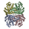
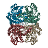
| Entry | Database: PDB / ID: 5x2z | ||||||
|---|---|---|---|---|---|---|---|
| Title | Crystal structure of Pseudomonas putida methionine gamma-lyase C116H mutant with L-methionine intermediates | ||||||
 Components Components | L-methionine gamma-lyase | ||||||
 Keywords Keywords |  LYASE / Pyridoxal 5'-phosphate LYASE / Pyridoxal 5'-phosphate | ||||||
| Function / homology |  Function and homology informationhomocysteine desulfhydrase / homocysteine desulfhydrase activity / methionine gamma-lyase / methionine gamma-lyase activity / transsulfuration / pyridoxal phosphate binding / cytoplasm Function and homology informationhomocysteine desulfhydrase / homocysteine desulfhydrase activity / methionine gamma-lyase / methionine gamma-lyase activity / transsulfuration / pyridoxal phosphate binding / cytoplasmSimilarity search - Function | ||||||
| Biological species |  Pseudomonas putida (bacteria) Pseudomonas putida (bacteria) | ||||||
| Method | X-RAY DIFFRACTION / SYNCHROTRON / MOLECULAR REPLACEMENT / Resolution: 1.8 Å | ||||||
 Authors Authors | Shiba, T. / Sato, D. / Harada, S. | ||||||
 Citation Citation | Journal: Protein Sci. / Year: 2017 Title: Structural and mechanistic insights into homocysteine degradation by a mutant of methionine gamma-lyase based on substrate-assisted catalysis Authors: Sato, D. / Shiba, T. / Yunoto, S. / Furutani, K. / Fukumoto, M. / Kudou, D. / Tamura, T. / Inagaki, K. / Harada, S. | ||||||
| History |
|
- Structure visualization
| Structure viewer | Molecule: MolmilJmol/JSmol |
|---|
- Downloads & links
-Download
| PDBx/mmCIF format | 5x2z.cif.gz | 318.4 KB | Display | PDBx/mmCIF format |
|---|---|---|---|---|
| PDB format | pdb5x2z.ent.gz | 259.1 KB | Display | PDB format |
| PDBx/mmJSON format | 5x2z.json.gz | Tree view | PDBx/mmJSON format | |
| Others |  Other downloads Other downloads |
-Validation report
| Arichive directory | https://data.pdbj.org/pub/pdb/validation_reports/x2/5x2zftp://data.pdbj.org/pub/pdb/validation_reports/x2/5x2z | HTTPS FTP |
|---|
-Related structure data
| Related structure data | 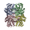 5x2vC  5x2wC 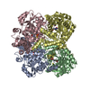 5x2xC  5x2yC  5x30C  2o7cS C: citing same article ( S: Starting model for refinement |
|---|---|
| Similar structure data |


-Links
 PDBj
PDBj- Assembly
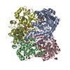
| Deposited unit | 
| ||||||||
|---|---|---|---|---|---|---|---|---|---|
| 1 |
| ||||||||
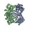
| Unit cell |
|
-Components
| #1: Protein | Mass: 42708.621 Da / Num. of mol.: 4 Source method: isolated from a genetically manipulated source Source: (gene. exp.) Pseudomonas putida (bacteria) / Gene: mdeA / Production host: Escherichia coli (E. coli)References: UniProt: P13254, methionine gamma-lyase, homocysteine desulfhydrase#2: Chemical | ChemComp-3LM / ( 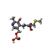  Mass: 378.338 Da / Num. of mol.: 4 / Source method: obtained synthetically / Formula: C13H19N2O7PS Mass: 378.338 Da / Num. of mol.: 4 / Source method: obtained synthetically / Formula: C13H19N2O7PS#3: Water | ChemComp-HOH / | Water Mass: 18.015 Da / Num. of mol.: 902 / Source method: isolated from a natural source / Formula: H2O Mass: 18.015 Da / Num. of mol.: 902 / Source method: isolated from a natural source / Formula: H2O |
|---|
-Experimental details
-Experiment
| Experiment | Method: X-RAY DIFFRACTION / Number of used crystals: 1 |
|---|
- Sample preparation
| Crystal | Density Matthews: 2.78 Å3/Da / Density % sol: 55.83 % |
|---|---|
| Crystal grow | Temperature: 293 K / Method: vapor diffusion, hanging drop Details: 0.2M Na-K phosphate buffer, 6-10% PEG 6000, 0.25M ammonium sulfate. 0.5mM PLP |
-Data collection
| Diffraction | Mean temperature: 100 K |
|---|---|
| Diffraction source | Source: SYNCHROTRON / Site: SPring-8  / Beamline: BL44XU / Wavelength: 0.9 Å / Beamline: BL44XU / Wavelength: 0.9 Å |
| Detector | Type: RAYONIX MX300HE / Detector: CCD / Date: Dec 17, 2013 |
| Radiation | Monochromator: Si 111 / Protocol: SINGLE WAVELENGTH / Monochromatic (M) / Laue (L): M / Scattering type: x-ray |
| Radiation wavelength | Wavelength: 0.9 Å / Relative weight: 1 |
| Reflection | Resolution: 1.8→50 Å / Num. obs: 176399 / % possible obs: 98.5 % / Redundancy: 4.4 % / Rmerge(I) obs: 0.102 / Net I/σ(I): 15.1 |
| Reflection shell | Resolution: 1.8→1.83 Å / Redundancy: 5.8 % / Rmerge(I) obs: 0.479 / Mean I/σ(I) obs: 2.4 / % possible all: 93.9 |
- Processing
| Software |
| ||||||||||||||||||||||||||||||||||||||||||||||||||||||||||||||||||||||||||||||||||||||||||||||||||||||||||||||||||||||||||||||||||||||||||||||||||||||||||||||||||||||||||||||||||||||
|---|---|---|---|---|---|---|---|---|---|---|---|---|---|---|---|---|---|---|---|---|---|---|---|---|---|---|---|---|---|---|---|---|---|---|---|---|---|---|---|---|---|---|---|---|---|---|---|---|---|---|---|---|---|---|---|---|---|---|---|---|---|---|---|---|---|---|---|---|---|---|---|---|---|---|---|---|---|---|---|---|---|---|---|---|---|---|---|---|---|---|---|---|---|---|---|---|---|---|---|---|---|---|---|---|---|---|---|---|---|---|---|---|---|---|---|---|---|---|---|---|---|---|---|---|---|---|---|---|---|---|---|---|---|---|---|---|---|---|---|---|---|---|---|---|---|---|---|---|---|---|---|---|---|---|---|---|---|---|---|---|---|---|---|---|---|---|---|---|---|---|---|---|---|---|---|---|---|---|---|---|---|---|---|
| Refinement | Method to determine structure: MOLECULAR REPLACEMENT Starting model: 2O7C Resolution: 1.8→20 Å / Cor.coef. Fo:Fc: 0.959 / Cor.coef. Fo:Fc free: 0.944 / SU B: 2.542 / SU ML: 0.076 / Cross valid method: THROUGHOUT / ESU R: 0.113 / ESU R Free: 0.108 / Details: HYDROGENS HAVE BEEN ADDED IN THE RIDING POSITIONS
| ||||||||||||||||||||||||||||||||||||||||||||||||||||||||||||||||||||||||||||||||||||||||||||||||||||||||||||||||||||||||||||||||||||||||||||||||||||||||||||||||||||||||||||||||||||||
| Solvent computation | Ion probe radii: 0.8 Å / Shrinkage radii: 0.8 Å / VDW probe radii: 1.2 Å | ||||||||||||||||||||||||||||||||||||||||||||||||||||||||||||||||||||||||||||||||||||||||||||||||||||||||||||||||||||||||||||||||||||||||||||||||||||||||||||||||||||||||||||||||||||||
| Displacement parameters | Biso mean: 26.913 Å2
| ||||||||||||||||||||||||||||||||||||||||||||||||||||||||||||||||||||||||||||||||||||||||||||||||||||||||||||||||||||||||||||||||||||||||||||||||||||||||||||||||||||||||||||||||||||||
| Refinement step | Cycle: 1 / Resolution: 1.8→20 Å
| ||||||||||||||||||||||||||||||||||||||||||||||||||||||||||||||||||||||||||||||||||||||||||||||||||||||||||||||||||||||||||||||||||||||||||||||||||||||||||||||||||||||||||||||||||||||
| Refine LS restraints |
|