 Movie
Movie Controller


+ Open data

- Basic information
| Entry | Database: PDB / ID: 5x17 | ||||||
|---|---|---|---|---|---|---|---|
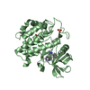
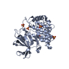
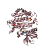
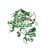
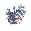
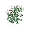
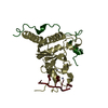
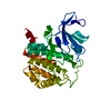
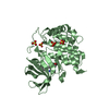
| Title | Crystal structure of murine CK1d in complex with ADP | ||||||
 Components Components | Casein kinase I isoform delta | ||||||
 Keywords Keywords |  TRANSFERASE / CK1D / KINASE TRANSFERASE / CK1D / KINASE | ||||||
| Function / homology |  Function and homology information Function and homology informationMajor pathway of rRNA processing in the nucleolus and cytosol / COPII-mediated vesicle transport / positive regulation of non-canonical Wnt signaling pathway / protein localization to Golgi apparatus / Loss of Nlp from mitotic centrosomes / Recruitment of mitotic centrosome proteins and complexes / Loss of proteins required for interphase microtubule organization from the centrosome / Recruitment of NuMA to mitotic centrosomes / Anchoring of the basal body to the plasma membrane / AURKA Activation by TPX2 ...Major pathway of rRNA processing in the nucleolus and cytosol / COPII-mediated vesicle transport / positive regulation of non-canonical Wnt signaling pathway / protein localization to Golgi apparatus / Loss of Nlp from mitotic centrosomes / Recruitment of mitotic centrosome proteins and complexes / Loss of proteins required for interphase microtubule organization from the centrosome / Recruitment of NuMA to mitotic centrosomes / Anchoring of the basal body to the plasma membrane / AURKA Activation by TPX2 / midbrain dopaminergic neuron differentiation / Regulation of PLK1 Activity at G2/M Transition / microtubule nucleation / protein localization to cilium / tau-protein kinase / non-motile cilium assembly / protein localization to centrosome / tau-protein kinase activity / Golgi organization / positive regulation of Wnt signaling pathway / spindle assembly / ciliary basal body / spindle microtubule / circadian regulation of gene expression / regulation of circadian rhythm / spindle / Wnt signaling pathway / endocytosis / positive regulation of canonical Wnt signaling pathway / positive regulation of proteasomal ubiquitin-dependent protein catabolic process / kinase activity / non-specific serine/threonine protein kinase / protein kinase activity / positive regulation of protein phosphorylation / protein phosphorylation / protein serine kinase activity / protein serine/threonine kinase activity / centrosome / perinuclear region of cytoplasm / Golgi apparatus / signal transduction / nucleoplasm / ATP binding / nucleus / plasma membrane / cytosolSimilarity search - Function | ||||||
| Biological species |  Mus musculus (house mouse) Mus musculus (house mouse) | ||||||
| Method | X-RAY DIFFRACTION / SYNCHROTRON / MOLECULAR REPLACEMENT / Resolution: 2 Å | ||||||
 Authors Authors | Kikuchi, M. / Shinohara, Y. / Ueda, H.R. / Umehara, T. | ||||||
 Citation Citation | Journal: Mol. Cell / Year: 2017 Title: Temperature-Sensitive Substrate and Product Binding Underlie Temperature-Compensated Phosphorylation in the Clock Authors: Shinohara, Y. / Koyama, Y.M. / Ukai-Tadenuma, M. / Hirokawa, T. / Kikuchi, M. / Yamada, R.G. / Ukai, H. / Fujishima, H. / Umehara, T. / Tainaka, K. / Ueda, H.R. | ||||||
| History |
|
- Structure visualization
| Structure viewer | Molecule: MolmilJmol/JSmol |
|---|
- Downloads & links
-Download
| PDBx/mmCIF format | 5x17.cif.gz | 139.4 KB | Display | PDBx/mmCIF format |
|---|---|---|---|---|
| PDB format | pdb5x17.ent.gz | 106.9 KB | Display | PDB format |
| PDBx/mmJSON format | 5x17.json.gz | Tree view | PDBx/mmJSON format | |
| Others |  Other downloads Other downloads |
-Validation report
| Arichive directory | https://data.pdbj.org/pub/pdb/validation_reports/x1/5x17ftp://data.pdbj.org/pub/pdb/validation_reports/x1/5x17 | HTTPS FTP |
|---|
-Related structure data
| Related structure data |  5x18C 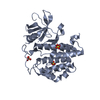 3uysS C: citing same article ( S: Starting model for refinement |
|---|---|
| Similar structure data |

-Links
 PDBj
PDBj


- Assembly
| Deposited unit | 
| ||||||||
|---|---|---|---|---|---|---|---|---|---|
| 1 | 
| ||||||||
| 2 | 
| ||||||||
| Unit cell |
|
-Components
| #1: Protein | Mass: 34320.637 Da / Num. of mol.: 2 / Fragment: UNP residues 1-294 Source method: isolated from a genetically manipulated source Source: (gene. exp.) Mus musculus (house mouse) / Gene: Csnk1d, Hckid / Plasmid: pET-28a(+) / Production host:  Escherichia coli (E. coli) / Strain (production host): Rosetta2(DE3) Escherichia coli (E. coli) / Strain (production host): Rosetta2(DE3)References: UniProt: Q9DC28, non-specific serine/threonine protein kinase, tau-protein kinase#2: Chemical | ChemComp-SO4 / Sulfate  Mass: 96.063 Da / Num. of mol.: 11 / Source method: obtained synthetically / Formula: SO4 Mass: 96.063 Da / Num. of mol.: 11 / Source method: obtained synthetically / Formula: SO4#3: Chemical | Adenosine diphosphate  Mass: 427.201 Da / Num. of mol.: 2 / Source method: obtained synthetically / Formula: C10H15N5O10P2 / Comment: ADP, energy-carrying molecule*YM Mass: 427.201 Da / Num. of mol.: 2 / Source method: obtained synthetically / Formula: C10H15N5O10P2 / Comment: ADP, energy-carrying molecule*YM#4: Water | ChemComp-HOH / | Water Mass: 18.015 Da / Num. of mol.: 311 / Source method: isolated from a natural source / Formula: H2O Mass: 18.015 Da / Num. of mol.: 311 / Source method: isolated from a natural source / Formula: H2O |
|---|
-Experimental details
-Experiment
| Experiment | Method: X-RAY DIFFRACTION / Number of used crystals: 1 |
|---|
- Sample preparation
| Crystal | Density Matthews: 2.29 Å3/Da / Density % sol: 46.19 % |
|---|---|
| Crystal grow | Temperature: 293 K / Method: vapor diffusion, sitting drop / pH: 5 Details: 100 mM Na-citrate, 25 mM (NH4)2SO4 and 7 % PEG 8000, pH 5.0 |
-Data collection
| Diffraction | Mean temperature: 100 K |
|---|---|
| Diffraction source | Source: SYNCHROTRON / Site: SPring-8  / Beamline: BL26B2 / Wavelength: 1 Å / Beamline: BL26B2 / Wavelength: 1 Å |
| Detector | Type: RAYONIX MX225HE / Detector: CCD / Date: Jul 22, 2016 |
| Radiation | Monochromator: Si double crystal / Protocol: SINGLE WAVELENGTH / Monochromatic (M) / Laue (L): M / Scattering type: x-ray |
| Radiation wavelength | Wavelength: 1 Å / Relative weight: 1 |
| Reflection | Resolution: 2→50 Å / Num. obs: 41687 / % possible obs: 100 % / Redundancy: 3.8 % / Net I/σ(I): 15.3 |
| Reflection shell | Resolution: 2→2.03 Å / Redundancy: 3.7 % / Rmerge(I) obs: 0.596 / Mean I/σ(I) obs: 2.2 / Num. unique obs: 2087 / % possible all: 99.9 |
- Processing
| Software |
| ||||||||||||||||||||||||||||||||||||||||||||||||||||||||||||||||||||||||||||||||||||||||||||||||||||||||||||||||||||||||||||||||||||||||||||||||||||||||||||||||||||||||||||||||||||||
|---|---|---|---|---|---|---|---|---|---|---|---|---|---|---|---|---|---|---|---|---|---|---|---|---|---|---|---|---|---|---|---|---|---|---|---|---|---|---|---|---|---|---|---|---|---|---|---|---|---|---|---|---|---|---|---|---|---|---|---|---|---|---|---|---|---|---|---|---|---|---|---|---|---|---|---|---|---|---|---|---|---|---|---|---|---|---|---|---|---|---|---|---|---|---|---|---|---|---|---|---|---|---|---|---|---|---|---|---|---|---|---|---|---|---|---|---|---|---|---|---|---|---|---|---|---|---|---|---|---|---|---|---|---|---|---|---|---|---|---|---|---|---|---|---|---|---|---|---|---|---|---|---|---|---|---|---|---|---|---|---|---|---|---|---|---|---|---|---|---|---|---|---|---|---|---|---|---|---|---|---|---|---|---|
| Refinement | Method to determine structure: MOLECULAR REPLACEMENT Starting model: 3UYS Resolution: 2→50 Å / Cor.coef. Fo:Fc: 0.956 / Cor.coef. Fo:Fc free: 0.923 / SU B: 5.38 / SU ML: 0.145 / Cross valid method: THROUGHOUT / ESU R: 0.196 / ESU R Free: 0.176 / Stereochemistry target values: MAXIMUM LIKELIHOOD / Details: HYDROGENS HAVE BEEN ADDED IN THE RIDING POSITIONS
| ||||||||||||||||||||||||||||||||||||||||||||||||||||||||||||||||||||||||||||||||||||||||||||||||||||||||||||||||||||||||||||||||||||||||||||||||||||||||||||||||||||||||||||||||||||||
| Solvent computation | Ion probe radii: 0.8 Å / Shrinkage radii: 0.8 Å / VDW probe radii: 1.4 Å / Solvent model: MASK | ||||||||||||||||||||||||||||||||||||||||||||||||||||||||||||||||||||||||||||||||||||||||||||||||||||||||||||||||||||||||||||||||||||||||||||||||||||||||||||||||||||||||||||||||||||||
| Displacement parameters | Biso mean: 36.415 Å2
| ||||||||||||||||||||||||||||||||||||||||||||||||||||||||||||||||||||||||||||||||||||||||||||||||||||||||||||||||||||||||||||||||||||||||||||||||||||||||||||||||||||||||||||||||||||||
| Refinement step | Cycle: 1 / Resolution: 2→50 Å
| ||||||||||||||||||||||||||||||||||||||||||||||||||||||||||||||||||||||||||||||||||||||||||||||||||||||||||||||||||||||||||||||||||||||||||||||||||||||||||||||||||||||||||||||||||||||
| Refine LS restraints |
|