 Movie
Movie Controller


+ Open data

- Basic information
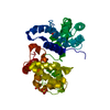
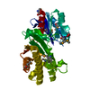
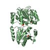
| Entry | Database: PDB / ID: 5wx8 | ||||||
|---|---|---|---|---|---|---|---|
| Title | Human herpesvirus 6A immediate early protein 2 C-terminal domain | ||||||
 Components Components | Immediate-early protein 2 | ||||||
 Keywords Keywords |  DNA BINDING PROTEIN / Transactivator DNA BINDING PROTEIN / Transactivator | ||||||
| Function / homology | Human herpesvirus 6 immediate early protein / Human herpesvirus 6 immediate early protein / Herpesvirus intermediate/early protein 2/3 / Herpes virus intermediate/early protein 2/3 / host cell nucleus / regulation of DNA-templated transcription / DNA binding / Immediate-early protein 2 Function and homology information Function and homology information | ||||||
| Biological species | Human herpesvirus 6A | ||||||
| Method | X-RAY DIFFRACTION / SYNCHROTRON / SAD / Resolution: 2.5 Å | ||||||
 Authors Authors | Nishimura, M. / Wang, J. / Wakata, A. / Sakamoto, K. / Mori, Y. | ||||||
 Citation Citation | Journal: J. Virol. / Year: 2017 Title: Crystal Structure of the DNA-Binding Domain of Human Herpesvirus 6A Immediate Early Protein 2. Authors: Nishimura, M. / Wang, J. / Wakata, A. / Sakamoto, K. / Mori, Y. | ||||||
| History |
|
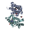
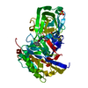
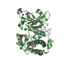
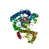
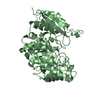
- Structure visualization
| Structure viewer | Molecule: MolmilJmol/JSmol |
|---|
- Downloads & links
-Download
| PDBx/mmCIF format | 5wx8.cif.gz | 143.8 KB | Display | PDBx/mmCIF format |
|---|---|---|---|---|
| PDB format | pdb5wx8.ent.gz | 120.3 KB | Display | PDB format |
| PDBx/mmJSON format | 5wx8.json.gz | Tree view | PDBx/mmJSON format | |
| Others |  Other downloads Other downloads |
-Validation report
| Arichive directory | https://data.pdbj.org/pub/pdb/validation_reports/wx/5wx8ftp://data.pdbj.org/pub/pdb/validation_reports/wx/5wx8 | HTTPS FTP |
|---|
-Related structure data
| Similar structure data |
|---|
-Links
 PDBj
PDBj- Assembly
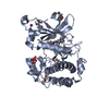
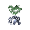
| Deposited unit | 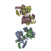
| ||||||||
|---|---|---|---|---|---|---|---|---|---|
| 1 | 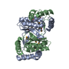
| ||||||||
| 2 | 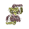
| ||||||||
| Unit cell |
|
-Components
| #1: Protein | Mass: 20833.508 Da / Num. of mol.: 4 / Fragment: C-terminal domain (UNP RESIDUES 1324-1500) Source method: isolated from a genetically manipulated source Source: (gene. exp.)  Human herpesvirus 6A (strain Uganda-1102) Human herpesvirus 6A (strain Uganda-1102)Strain: Uganda-1102 / Gene: U90/U87/U86, BCLF0 / Production host:  Escherichia coli (E. coli) / References: UniProt: Q77Z83 Escherichia coli (E. coli) / References: UniProt: Q77Z83#2: Water | ChemComp-HOH / | Water Mass: 18.015 Da / Num. of mol.: 298 / Source method: isolated from a natural source / Formula: H2O Mass: 18.015 Da / Num. of mol.: 298 / Source method: isolated from a natural source / Formula: H2O |
|---|
-Experimental details
-Experiment
| Experiment | Method: X-RAY DIFFRACTION / Number of used crystals: 1 |
|---|
- Sample preparation
| Crystal | Density Matthews: 2.38 Å3/Da / Density % sol: 48.23 % |
|---|---|
| Crystal grow | Temperature: 277 K / Method: vapor diffusion, sitting drop / pH: 8.8 Details: 24% polyethylene glycol 3350, 0.2M sodium formate, 20 mM Tris-HCl (pH 8.8), 20 mM CaCl2, 1 mM DTT |
-Data collection
| Diffraction | Mean temperature: 100 K |
|---|---|
| Diffraction source | Source: SYNCHROTRON / Site: SPring-8  / Beamline: BL38B1 / Wavelength: 0.97905 Å / Beamline: BL38B1 / Wavelength: 0.97905 Å |
| Detector | Type: RAYONIX MX225HE / Detector: CCD / Date: Jul 23, 2016 |
| Radiation | Protocol: SINGLE WAVELENGTH / Monochromatic (M) / Laue (L): M / Scattering type: x-ray |
| Radiation wavelength | Wavelength: 0.97905 Å / Relative weight: 1 |
| Reflection | Resolution: 2.5→42.51 Å / Num. obs: 27197 / % possible obs: 97.88 % / Redundancy: 6.3 % / Net I/σ(I): 9.47 |
- Processing
| Software |
| |||||||||||||||||||||||||||||||||||||||||||||||||||||||||||||||||||||||||||||||||||||||||||||||||||||||||
|---|---|---|---|---|---|---|---|---|---|---|---|---|---|---|---|---|---|---|---|---|---|---|---|---|---|---|---|---|---|---|---|---|---|---|---|---|---|---|---|---|---|---|---|---|---|---|---|---|---|---|---|---|---|---|---|---|---|---|---|---|---|---|---|---|---|---|---|---|---|---|---|---|---|---|---|---|---|---|---|---|---|---|---|---|---|---|---|---|---|---|---|---|---|---|---|---|---|---|---|---|---|---|---|---|---|---|
| Refinement | Method to determine structure: SAD / Resolution: 2.5→42.508 Å / SU ML: 0.33 / Cross valid method: FREE R-VALUE / σ(F): 1.5 / Phase error: 26 / Stereochemistry target values: MLHL
| |||||||||||||||||||||||||||||||||||||||||||||||||||||||||||||||||||||||||||||||||||||||||||||||||||||||||
| Solvent computation | Shrinkage radii: 0.9 Å / VDW probe radii: 1.11 Å / Solvent model: FLAT BULK SOLVENT MODEL | |||||||||||||||||||||||||||||||||||||||||||||||||||||||||||||||||||||||||||||||||||||||||||||||||||||||||
| Refinement step | Cycle: LAST / Resolution: 2.5→42.508 Å
| |||||||||||||||||||||||||||||||||||||||||||||||||||||||||||||||||||||||||||||||||||||||||||||||||||||||||
| Refine LS restraints |
| |||||||||||||||||||||||||||||||||||||||||||||||||||||||||||||||||||||||||||||||||||||||||||||||||||||||||
| LS refinement shell |
|
