 Movie
Movie Controller

[English] 日本語
 Yorodumi
- PDB-5wvo: Crystal structure of DNMT1 RFTS domain in complex with K18/K23 mo... -
+ Open data

- Basic information
| Entry | Database: PDB / ID: 5wvo | ||||||
|---|---|---|---|---|---|---|---|
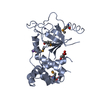
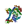
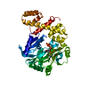
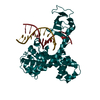
| Title | Crystal structure of DNMT1 RFTS domain in complex with K18/K23 mono-ubiquitylated histone H3 | ||||||
 Components Components |
| ||||||
 Keywords Keywords |  SIGNALING PROTEIN/TRANSFERASE / DNA methylation / Ubiquitination / SIGNALING PROTEIN-TRANSFERASE complex SIGNALING PROTEIN/TRANSFERASE / DNA methylation / Ubiquitination / SIGNALING PROTEIN-TRANSFERASE complex | ||||||
| Function / homology |  Function and homology information Function and homology informationchromosomal DNA methylation maintenance following DNA replication / negative regulation of vascular associated smooth muscle cell differentiation involved in phenotypic switching / epigenetic programming of gene expression / cellular response to bisphenol A / negative regulation of vascular associated smooth muscle cell apoptotic process / DNA-methyltransferase activity / DNA (cytosine-5-)-methyltransferase / DNA (cytosine-5-)-methyltransferase activity / SUMOylation of DNA methylation proteins / DNA methylation-dependent heterochromatin formation ...chromosomal DNA methylation maintenance following DNA replication / negative regulation of vascular associated smooth muscle cell differentiation involved in phenotypic switching / epigenetic programming of gene expression / cellular response to bisphenol A / negative regulation of vascular associated smooth muscle cell apoptotic process / DNA-methyltransferase activity / DNA (cytosine-5-)-methyltransferase / DNA (cytosine-5-)-methyltransferase activity / SUMOylation of DNA methylation proteins / DNA methylation-dependent heterochromatin formation / STAT3 nuclear events downstream of ALK signaling / female germ cell nucleus / methyl-CpG binding / negative regulation of gene expression via chromosomal CpG island methylation / Formation of the ternary complex, and subsequently, the 43S complex / Ribosomal scanning and start codon recognition / Translation initiation complex formation / SARS-CoV-1 modulates host translation machinery / Peptide chain elongation / Selenocysteine synthesis / Formation of a pool of free 40S subunits / Eukaryotic Translation Termination / Response of EIF2AK4 (GCN2) to amino acid deficiency / SRP-dependent cotranslational protein targeting to membrane / Viral mRNA Translation / Nonsense Mediated Decay (NMD) independent of the Exon Junction Complex (EJC) / GTP hydrolysis and joining of the 60S ribosomal subunit / L13a-mediated translational silencing of Ceruloplasmin expression / Major pathway of rRNA processing in the nucleolus and cytosol / Nuclear events stimulated by ALK signaling in cancer / Nonsense Mediated Decay (NMD) enhanced by the Exon Junction Complex (EJC) / pericentric heterochromatin / Chromatin modifying enzymes / epigenetic regulation of gene expression / Maturation of protein E / Maturation of protein E / cytosolic ribosome / ER Quality Control Compartment (ERQC) / Myoclonic epilepsy of Lafora / FLT3 signaling by CBL mutants / Prevention of phagosomal-lysosomal fusion / IRAK2 mediated activation of TAK1 complex / Alpha-protein kinase 1 signaling pathway / Glycogen synthesis / positive regulation of vascular associated smooth muscle cell proliferation / IRAK1 recruits IKK complex / IRAK1 recruits IKK complex upon TLR7/8 or 9 stimulation / Membrane binding and targetting of GAG proteins / Endosomal Sorting Complex Required For Transport (ESCRT) / IRAK2 mediated activation of TAK1 complex upon TLR7/8 or 9 stimulation / PTK6 Regulates RTKs and Their Effectors AKT1 and DOK1 / Negative regulation of FLT3 / Constitutive Signaling by NOTCH1 HD Domain Mutants / Regulation of FZD by ubiquitination / TICAM1,TRAF6-dependent induction of TAK1 complex / NOTCH2 Activation and Transmission of Signal to the Nucleus / TICAM1-dependent activation of IRF3/IRF7 / APC/C:Cdc20 mediated degradation of Cyclin B / p75NTR recruits signalling complexes / Downregulation of ERBB4 signaling / TRAF6 mediated IRF7 activation in TLR7/8 or 9 signaling / APC-Cdc20 mediated degradation of Nek2A / PINK1-PRKN Mediated Mitophagy / telomere organization / TRAF6-mediated induction of TAK1 complex within TLR4 complex / Pexophagy / Regulation of innate immune responses to cytosolic DNA / VLDLR internalisation and degradation / InlA-mediated entry of Listeria monocytogenes into host cells / Downregulation of ERBB2:ERBB3 signaling / RNA Polymerase I Promoter Opening / NF-kB is activated and signals survival / Interleukin-7 signaling / NRIF signals cell death from the nucleus / Regulation of PTEN localization / Activated NOTCH1 Transmits Signal to the Nucleus / Regulation of BACH1 activity / Assembly of the ORC complex at the origin of replication / Translesion synthesis by REV1 / Synthesis of active ubiquitin: roles of E1 and E2 enzymes / Translesion synthesis by POLK / MAP3K8 (TPL2)-dependent MAPK1/3 activation / TICAM1, RIP1-mediated IKK complex recruitment / Downregulation of TGF-beta receptor signaling / Activation of IRF3, IRF7 mediated by TBK1, IKKε (IKBKE) / Translesion synthesis by POLI / Gap-filling DNA repair synthesis and ligation in GG-NER / Josephin domain DUBs / DNA methylation / Regulation of activated PAK-2p34 by proteasome mediated degradation / InlB-mediated entry of Listeria monocytogenes into host cell / IKK complex recruitment mediated by RIP1 / JNK (c-Jun kinases) phosphorylation and activation mediated by activated human TAK1 / Condensation of Prophase Chromosomes / ERCC6 (CSB) and EHMT2 (G9a) positively regulate rRNA expression / SIRT1 negatively regulates rRNA expression / Chromatin modifications during the maternal to zygotic transition (MZT) / HCMV Late Events / TGF-beta receptor signaling in EMT (epithelial to mesenchymal transition) / N-glycan trimming in the ER and Calnexin/Calreticulin cycleSimilarity search - Function | ||||||
| Biological species |  Homo sapiens (human) Homo sapiens (human) | ||||||
| Method | X-RAY DIFFRACTION / SYNCHROTRON / MOLECULAR REPLACEMENT / Resolution: 1.997 Å | ||||||
 Authors Authors | Ishiyama, S. / Nishiyama, A. / Nakanishi, M. / Arita, K. | ||||||
| Funding support |  Japan, 1items Japan, 1items
| ||||||
 Citation Citation | Journal: Mol. Cell / Year: 2017 Title: Structure of the Dnmt1 Reader Module Complexed with a Unique Two-Mono-Ubiquitin Mark on Histone H3 Reveals the Basis for DNA Methylation Maintenance Authors: Ishiyama, S. / Nishiyama, A. / Saeki, Y. / Moritsugu, K. / Morimoto, D. / Yamaguchi, L. / Arai, N. / Matsumura, R. / Kawakami, T. / Mishima, Y. / Hojo, H. / Shimamura, S. / Ishikawa, F. / ...Authors: Ishiyama, S. / Nishiyama, A. / Saeki, Y. / Moritsugu, K. / Morimoto, D. / Yamaguchi, L. / Arai, N. / Matsumura, R. / Kawakami, T. / Mishima, Y. / Hojo, H. / Shimamura, S. / Ishikawa, F. / Tajima, S. / Tanaka, K. / Ariyoshi, M. / Shirakawa, M. / Ikeguchi, M. / Kidera, A. / Suetake, I. / Arita, K. / Nakanishi, M. | ||||||
| History |
|
- Structure visualization
| Structure viewer | Molecule: MolmilJmol/JSmol |
|---|
- Downloads & links
-Download
| PDBx/mmCIF format | 5wvo.cif.gz | 99 KB | Display | PDBx/mmCIF format |
|---|---|---|---|---|
| PDB format | pdb5wvo.ent.gz | 73.2 KB | Display | PDB format |
| PDBx/mmJSON format | 5wvo.json.gz | Tree view | PDBx/mmJSON format | |
| Others |  Other downloads Other downloads |
-Validation report
| Arichive directory | https://data.pdbj.org/pub/pdb/validation_reports/wv/5wvoftp://data.pdbj.org/pub/pdb/validation_reports/wv/5wvo | HTTPS FTP |
|---|
-Related structure data


-Links
 PDBj
PDBj


- Assembly
| Deposited unit | 
| ||||||||
|---|---|---|---|---|---|---|---|---|---|
| 1 |
| ||||||||
| Unit cell |
|
-Components
| #1: Protein | Mass: 8622.922 Da / Num. of mol.: 2 / Mutation: G76C Source method: isolated from a genetically manipulated source Source: (gene. exp.) Homo sapiens (human) / Gene: RPS27A, UBA80, UBCEP1 / Plasmid: pET22bProduction host: Strain (production host): BL21(DE3) / References: UniProt: P62979 #2: Protein | | Mass: 27980.334 Da / Num. of mol.: 1 / Fragment: RFTS domain, UNP residues 351-600 Source method: isolated from a genetically manipulated source Source: (gene. exp.) Homo sapiens (human) / Gene: DNMT1, AIM, CXXC9, DNMT / Plasmid: modified pGEX4T vectorProduction host: Strain (production host): Rosetta2 (DE3) References: UniProt: P26358, DNA (cytosine-5-)-methyltransferase#3: Protein/peptide | | Histone H3 / Histone H3/a / Histone H3/b / Histone H3/c / Histone H3/d / Histone H3/f / Histone H3/h / Histone ...Histone H3/a / Histone H3/b / Histone H3/c / Histone H3/d / Histone H3/f / Histone H3/h / Histone H3/i / Histone H3/j / Histone H3/k / Histone H3/lMass: 3824.465 Da / Num. of mol.: 1 / Fragment: UNP residues 2-37 / Mutation: K18C,K23C Source method: isolated from a genetically manipulated source Source: (gene. exp.) Homo sapiens (human)Gene: HIST1H3A, H3FA, HIST1H3B, H3FL, HIST1H3C, H3FC, HIST1H3D, H3FB, HIST1H3E, H3FD, HIST1H3F, H3FI, HIST1H3G, H3FH, HIST1H3H, H3FK, HIST1H3I, H3FF, HIST1H3J, H3FJ Plasmid: modified pGEX4T Production host: Strain (production host): BL21(DE3) / References: UniProt: P68431 #4: Chemical | ChemComp-ZN / |   Mass: 65.409 Da / Num. of mol.: 1 / Source method: obtained synthetically / Formula: Zn Mass: 65.409 Da / Num. of mol.: 1 / Source method: obtained synthetically / Formula: Zn#5: Water | ChemComp-HOH / | Water Mass: 18.015 Da / Num. of mol.: 197 / Source method: isolated from a natural source / Formula: H2O Mass: 18.015 Da / Num. of mol.: 197 / Source method: isolated from a natural source / Formula: H2O |
|---|
-Experimental details
-Experiment
| Experiment | Method: X-RAY DIFFRACTION / Number of used crystals: 1 |
|---|
- Sample preparation
| Crystal | Density Matthews: 2.89 Å3/Da / Density % sol: 57.43 % |
|---|---|
| Crystal grow | Temperature: 277 K / Method: vapor diffusion, hanging drop / pH: 6 Details: 100mM Bis-Tris (pH 6.0), 200mM lithium sulfate monohydrate, 20% PEG 10000 |
-Data collection
| Diffraction | Mean temperature: 93 K |
|---|---|
| Diffraction source | Source: SYNCHROTRON / Site: Photon Factory / Beamline: BL-5A / Wavelength: 1.1 Å |
| Detector | Type: DECTRIS PILATUS 2M-F / Detector: PIXEL / Date: Nov 8, 2016 |
| Radiation | Protocol: SINGLE WAVELENGTH / Monochromatic (M) / Laue (L): M / Scattering type: x-ray |
| Radiation wavelength | Wavelength: 1.1 Å / Relative weight: 1 |
| Reflection | Resolution: 1.997→34.181 Å / Num. obs: 35892 / % possible obs: 99.8 % / Redundancy: 6.4 % / Net I/σ(I): 6.7 |
| Reflection shell | Resolution: 2→2.03 Å / Redundancy: 6.1 % / Rmerge(I) obs: 0.706 / Mean I/σ(I) obs: 3.6 / CC1/2: 0.778 / % possible all: 99.1 |
- Processing
| Software |
| ||||||||||||||||||||||||||||||||||||||||||||||||||||||||||||||||||||||||||||||||||||||||||||||||||
|---|---|---|---|---|---|---|---|---|---|---|---|---|---|---|---|---|---|---|---|---|---|---|---|---|---|---|---|---|---|---|---|---|---|---|---|---|---|---|---|---|---|---|---|---|---|---|---|---|---|---|---|---|---|---|---|---|---|---|---|---|---|---|---|---|---|---|---|---|---|---|---|---|---|---|---|---|---|---|---|---|---|---|---|---|---|---|---|---|---|---|---|---|---|---|---|---|---|---|---|
| Refinement | Method to determine structure: MOLECULAR REPLACEMENT Starting model: 1UBQ, 3EPZ Resolution: 1.997→34.181 Å / SU ML: 0.22 / Cross valid method: FREE R-VALUE / σ(F): 1.36 / Phase error: 23.85
| ||||||||||||||||||||||||||||||||||||||||||||||||||||||||||||||||||||||||||||||||||||||||||||||||||
| Solvent computation | Shrinkage radii: 0.9 Å / VDW probe radii: 1.11 Å | ||||||||||||||||||||||||||||||||||||||||||||||||||||||||||||||||||||||||||||||||||||||||||||||||||
| Refinement step | Cycle: LAST / Resolution: 1.997→34.181 Å
| ||||||||||||||||||||||||||||||||||||||||||||||||||||||||||||||||||||||||||||||||||||||||||||||||||
| Refine LS restraints |
| ||||||||||||||||||||||||||||||||||||||||||||||||||||||||||||||||||||||||||||||||||||||||||||||||||
| LS refinement shell |
|
