 Movie
Movie Controller

[English] 日本語
 Yorodumi
- PDB-5wia: Crystal structure of the segment, GNNSYS, from the low complexity... -
+ Open data

- Basic information
| Entry | Database: PDB / ID: 5wia | ||||||
|---|---|---|---|---|---|---|---|
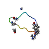
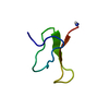
| Title | Crystal structure of the segment, GNNSYS, from the low complexity domain of TDP-43, residues 370-375 | ||||||
 Components Components | TAR DNA-binding protein 43 | ||||||
 Keywords Keywords | PROTEIN FIBRIL / Amyloid / TDP-43 | ||||||
| Function / homology |  Function and homology information Function and homology informationnuclear inner membrane organization / interchromatin granule / perichromatin fibrils / 3'-UTR-mediated mRNA destabilization / 3'-UTR-mediated mRNA stabilization / intracellular non-membrane-bounded organelle / negative regulation by host of viral transcription / pre-mRNA intronic binding / response to endoplasmic reticulum stress / RNA splicing ...nuclear inner membrane organization / interchromatin granule / perichromatin fibrils / 3'-UTR-mediated mRNA destabilization / 3'-UTR-mediated mRNA stabilization / intracellular non-membrane-bounded organelle / negative regulation by host of viral transcription / pre-mRNA intronic binding / response to endoplasmic reticulum stress / RNA splicing / negative regulation of protein phosphorylation / molecular condensate scaffold activity / mRNA 3'-UTR binding / regulation of protein stability / regulation of circadian rhythm / positive regulation of insulin secretion / mRNA processing / cytoplasmic stress granule / positive regulation of protein import into nucleus / rhythmic process / regulation of gene expression / double-stranded DNA binding / regulation of apoptotic process / amyloid fibril formation / regulation of cell cycle / nuclear speck / RNA polymerase II cis-regulatory region sequence-specific DNA binding / negative regulation of gene expression / lipid binding / mitochondrion / DNA binding / RNA binding / nucleoplasm / identical protein binding / nucleusSimilarity search - Function | ||||||
| Biological species |  Homo sapiens (human) Homo sapiens (human) | ||||||
| Method | X-RAY DIFFRACTION / SYNCHROTRON / MOLECULAR REPLACEMENT / Resolution: 1.002 Å | ||||||
 Authors Authors | Guenther, E.L. / Trinh, H. / Sawaya, M.R. / Eisenberg, D.S. | ||||||
| Funding support |  United States, 1items United States, 1items
| ||||||
 Citation Citation | Journal: Nat Struct Mol Biol / Year: 2018 Title: Atomic structures of TDP-43 LCD segments and insights into reversible or pathogenic aggregation. Authors: Elizabeth L Guenther / Qin Cao / Hamilton Trinh / Jiahui Lu / Michael R Sawaya / Duilio Cascio / David R Boyer / Jose A Rodriguez / Michael P Hughes / David S Eisenberg / Abstract: The normally soluble TAR DNA-binding protein 43 (TDP-43) is found aggregated both in reversible stress granules and in irreversible pathogenic amyloid. In TDP-43, the low-complexity domain (LCD) is ...The normally soluble TAR DNA-binding protein 43 (TDP-43) is found aggregated both in reversible stress granules and in irreversible pathogenic amyloid. In TDP-43, the low-complexity domain (LCD) is believed to be involved in both types of aggregation. To uncover the structural origins of these two modes of β-sheet-rich aggregation, we have determined ten structures of segments of the LCD of human TDP-43. Six of these segments form steric zippers characteristic of the spines of pathogenic amyloid fibrils; four others form LARKS, the labile amyloid-like interactions characteristic of protein hydrogels and proteins found in membraneless organelles, including stress granules. Supporting a hypothetical pathway from reversible to irreversible amyloid aggregation, we found that familial ALS variants of TDP-43 convert LARKS to irreversible aggregates. Our structures suggest how TDP-43 adopts both reversible and irreversible β-sheet aggregates and the role of mutation in the possible transition of reversible to irreversible pathogenic aggregation. | ||||||
| History |
|
- Structure visualization
| Structure viewer | Molecule: MolmilJmol/JSmol |
|---|
- Downloads & links
-Download
| PDBx/mmCIF format | 5wia.cif.gz | 10.7 KB | Display | PDBx/mmCIF format |
|---|---|---|---|---|
| PDB format | pdb5wia.ent.gz | 5.5 KB | Display | PDB format |
| PDBx/mmJSON format | 5wia.json.gz | Tree view | PDBx/mmJSON format | |
| Others |  Other downloads Other downloads |
-Validation report
| Arichive directory | https://data.pdbj.org/pub/pdb/validation_reports/wi/5wiaftp://data.pdbj.org/pub/pdb/validation_reports/wi/5wia | HTTPS FTP |
|---|
-Related structure data
| Related structure data |  7466C  7467C  8857C  5whnC  5whpC 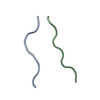 5wiqC  5wkbC  5wkdC  6cb9C  6cewC 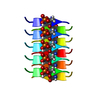 6cf4C  6cfhC C: citing same article ( |
|---|---|
| Similar structure data |


-Links
 PDBj
PDBj
- Assembly
| Deposited unit | 
| ||||||||
|---|---|---|---|---|---|---|---|---|---|
| 1 | x 10
| ||||||||
| Unit cell |
|
-Components
| #1: Protein/peptide | / TDP-43 Mass: 640.602 Da / Num. of mol.: 1 / Fragment: UNP residues 370-375 / Source method: obtained synthetically Details: Synthetic peptide GNNSYS corresponding to segment 370-375 of TDP-43 Source: (synth.) Homo sapiens (human) / References: UniProt: Q13148 |
|---|---|
| #2: Water | ChemComp-HOH / Water Mass: 18.015 Da / Num. of mol.: 1 / Source method: isolated from a natural source / Formula: H2O Mass: 18.015 Da / Num. of mol.: 1 / Source method: isolated from a natural source / Formula: H2O |
-Experimental details
-Experiment
| Experiment | Method: X-RAY DIFFRACTION / Number of used crystals: 1 |
|---|
- Sample preparation
| Crystal grow | Temperature: 298 K / Method: vapor diffusion, hanging drop / pH: 8.5 Details: 100mM bis tris propane 8.5, 200mM sodium nitrate, 20% PEG 3350 |
|---|
-Data collection
| Diffraction | Mean temperature: 100 K | |||||||||||||||||||||||||||||||||||||||||||||||||||||||||||||||||||||||||||||||||||||||||||||||||||
|---|---|---|---|---|---|---|---|---|---|---|---|---|---|---|---|---|---|---|---|---|---|---|---|---|---|---|---|---|---|---|---|---|---|---|---|---|---|---|---|---|---|---|---|---|---|---|---|---|---|---|---|---|---|---|---|---|---|---|---|---|---|---|---|---|---|---|---|---|---|---|---|---|---|---|---|---|---|---|---|---|---|---|---|---|---|---|---|---|---|---|---|---|---|---|---|---|---|---|---|---|
| Diffraction source | Source: SYNCHROTRON / Site: APS / Beamline: 24-ID-E / Wavelength: 0.9791 Å | |||||||||||||||||||||||||||||||||||||||||||||||||||||||||||||||||||||||||||||||||||||||||||||||||||
| Detector | Type: ADSC QUANTUM 315 / Detector: CCD / Date: Apr 18, 2015 | |||||||||||||||||||||||||||||||||||||||||||||||||||||||||||||||||||||||||||||||||||||||||||||||||||
| Radiation | Protocol: SINGLE WAVELENGTH / Monochromatic (M) / Laue (L): M / Scattering type: x-ray | |||||||||||||||||||||||||||||||||||||||||||||||||||||||||||||||||||||||||||||||||||||||||||||||||||
| Radiation wavelength | Wavelength: 0.9791 Å / Relative weight: 1 | |||||||||||||||||||||||||||||||||||||||||||||||||||||||||||||||||||||||||||||||||||||||||||||||||||
| Reflection | Resolution: 1→100 Å / Num. obs: 1864 / % possible obs: 95.2 % / Redundancy: 5.4 % / Biso Wilson estimate: 2.69 Å2 / Rmerge(I) obs: 0.155 / Rpim(I) all: 0.072 / Rrim(I) all: 0.172 / Χ2: 1.191 / Net I/σ(I): 5.2 / Num. measured all: 10032 | |||||||||||||||||||||||||||||||||||||||||||||||||||||||||||||||||||||||||||||||||||||||||||||||||||
| Reflection shell | Diffraction-ID: 1
|
- Processing
| Software |
| ||||||||||||||||||||||||
|---|---|---|---|---|---|---|---|---|---|---|---|---|---|---|---|---|---|---|---|---|---|---|---|---|---|
| Refinement | Method to determine structure: MOLECULAR REPLACEMENT Starting model: ideal 5-residue polyalanine beta-strand Resolution: 1.002→20.34 Å / SU ML: 0.07 / Cross valid method: FREE R-VALUE / σ(F): 1.39 / Phase error: 17.13
| ||||||||||||||||||||||||
| Solvent computation | Shrinkage radii: 0.9 Å / VDW probe radii: 1.11 Å | ||||||||||||||||||||||||
| Displacement parameters | Biso max: 15.34 Å2 / Biso mean: 3.7159 Å2 / Biso min: 1.17 Å2 | ||||||||||||||||||||||||
| Refinement step | Cycle: final / Resolution: 1.002→20.34 Å
| ||||||||||||||||||||||||
| Refine LS restraints |
| ||||||||||||||||||||||||
| LS refinement shell | Resolution: 1.0016→20.3437 Å / Rfactor Rfree error: 0 / Total num. of bins used: 1
|
