 Movie
Movie Controller

[English] 日本語
 Yorodumi
- PDB-5wg8: Structure of PP5C with LB-100; 7-oxabicyclo[2.2.1]heptane-2,3-dic... -
+ Open data

- Basic information
| Entry | Database: PDB / ID: 5wg8 | ||||||
|---|---|---|---|---|---|---|---|
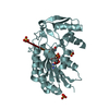
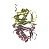
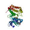
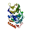
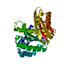
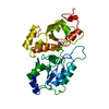
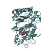
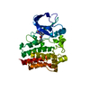
| Title | Structure of PP5C with LB-100; 7-oxabicyclo[2.2.1]heptane-2,3-dicarbonyl moiety modeled in the density | ||||||
 Components Components | Serine/threonine-protein phosphatase 5 | ||||||
 Keywords Keywords | HYDROLASE/HYDROLASE inhibitor / Hydrolase Inhibitor /  HYDROLASE / HYDROLASE-HYDROLASE inhibitor complex HYDROLASE / HYDROLASE-HYDROLASE inhibitor complex | ||||||
| Function / homology |  Function and homology information Function and homology informationresponse to arachidonic acid / peptidyl-serine dephosphorylation / peptidyl-threonine dephosphorylation / response to morphine / protein folding chaperone complex / myosin phosphatase activity / protein serine/threonine phosphatase activity / protein-serine/threonine phosphatase / phosphatase activity / phosphoprotein phosphatase activity ...response to arachidonic acid / peptidyl-serine dephosphorylation / peptidyl-threonine dephosphorylation / response to morphine / protein folding chaperone complex / myosin phosphatase activity / protein serine/threonine phosphatase activity / protein-serine/threonine phosphatase / phosphatase activity / phosphoprotein phosphatase activity / protein dephosphorylation / ESR-mediated signaling / response to lead ion / Hsp90 protein binding / tau protein binding / ADP binding / Negative regulation of MAPK pathway / MAPK cascade / double-strand break repair / Recruitment and ATM-mediated phosphorylation of repair and signaling proteins at DNA double strand breaks / mitotic cell cycle / positive regulation of canonical NF-kappaB signal transduction / intracellular membrane-bounded organelle / DNA-templated transcription / lipid binding / protein-containing complex / RNA binding / nucleoplasm / ATP binding / identical protein binding / metal ion binding / nucleus / plasma membrane / cytosolSimilarity search - Function | ||||||
| Biological species |  Homo sapiens (human) Homo sapiens (human) | ||||||
| Method | X-RAY DIFFRACTION / MOLECULAR REPLACEMENT / Resolution: 1.65 Å | ||||||
 Authors Authors | D'Arcy, B.M. / Swingle, M.R. / Honkanen, R.E. / Prakash, A. | ||||||
 Citation Citation | Journal: Mol. Cancer Ther. / Year: 2019 Title: The Antitumor Drug LB-100 Is a Catalytic Inhibitor of Protein Phosphatase 2A (PPP2CA) and 5 (PPP5C) Coordinating with the Active-Site Catalytic Metals in PPP5C. Authors: D'Arcy, B.M. / Swingle, M.R. / Papke, C.M. / Abney, K.A. / Bouska, E.S. / Prakash, A. / Honkanen, R.E. | ||||||
| History |
|
- Structure visualization
| Structure viewer | Molecule: MolmilJmol/JSmol |
|---|
- Downloads & links
-Download
| PDBx/mmCIF format | 5wg8.cif.gz | 150.2 KB | Display | PDBx/mmCIF format |
|---|---|---|---|---|
| PDB format | pdb5wg8.ent.gz | 113.3 KB | Display | PDB format |
| PDBx/mmJSON format | 5wg8.json.gz | Tree view | PDBx/mmJSON format | |
| Others |  Other downloads Other downloads |
-Validation report
| Arichive directory | https://data.pdbj.org/pub/pdb/validation_reports/wg/5wg8ftp://data.pdbj.org/pub/pdb/validation_reports/wg/5wg8 | HTTPS FTP |
|---|
-Related structure data
| Related structure data | 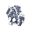 1s95S S: Starting model for refinement |
|---|---|
| Similar structure data |


-Links
 PDBj
PDBj


- Assembly
| Deposited unit | 
| ||||||||
|---|---|---|---|---|---|---|---|---|---|
| 1 |
| ||||||||
| Unit cell |
|
-Components
-Protein , 1 types, 1 molecules A
| #1: Protein | Mass: 37887.020 Da / Num. of mol.: 1 / Fragment: UNP residues 169-499 Source method: isolated from a genetically manipulated source Source: (gene. exp.) Homo sapiens (human) / Gene: PPP5C, PPP5 / Production host:  Escherichia coli (E. coli) Escherichia coli (E. coli)References: UniProt: P53041, protein-serine/threonine phosphatase |
|---|
-Non-polymers , 5 types, 267 molecules 


| #2: Chemical |  Mass: 54.938 Da / Num. of mol.: 2 / Source method: obtained synthetically / Formula: Mn Mass: 54.938 Da / Num. of mol.: 2 / Source method: obtained synthetically / Formula: Mn#3: Chemical | ChemComp-LB1 / ( |  Mass: 268.309 Da / Num. of mol.: 1 / Source method: obtained synthetically / Formula: C13H20N2O4 / Feature type: SUBJECT OF INVESTIGATION Mass: 268.309 Da / Num. of mol.: 1 / Source method: obtained synthetically / Formula: C13H20N2O4 / Feature type: SUBJECT OF INVESTIGATION#4: Chemical | ChemComp-MPD / ( | 2-Methyl-2,4-pentanediol Mass: 118.174 Da / Num. of mol.: 1 / Source method: obtained synthetically / Formula: C6H14O2 / Comment: precipitant*YM Mass: 118.174 Da / Num. of mol.: 1 / Source method: obtained synthetically / Formula: C6H14O2 / Comment: precipitant*YM#5: Chemical | ChemComp-MRD / ( | 2-Methyl-2,4-pentanediol Mass: 118.174 Da / Num. of mol.: 1 / Source method: obtained synthetically / Formula: C6H14O2 / Comment: precipitant*YM Mass: 118.174 Da / Num. of mol.: 1 / Source method: obtained synthetically / Formula: C6H14O2 / Comment: precipitant*YM#6: Water | ChemComp-HOH / | WaterMass: 18.015 Da / Num. of mol.: 262 / Source method: isolated from a natural source / Formula: H2O |
|---|
-Experimental details
-Experiment
| Experiment | Method: X-RAY DIFFRACTION / Number of used crystals: 1 |
|---|
- Sample preparation
| Crystal | Density Matthews: 2.43 Å3/Da / Density % sol: 49.4 % |
|---|---|
| Crystal grow | Temperature: 289.15 K / Method: vapor diffusion, sitting drop / pH: 8 Details: 10 mM Tris-HCl pH 8.0, 35% 2-methyl-2,4-pentanediol, and 10% polyethylene glycol methyl ether 5000 |
-Data collection
| Diffraction | Mean temperature: 100 K |
|---|---|
| Diffraction source | Source: SEALED TUBE / Type: BRUKER IMUS MICROFOCUS / Wavelength: 1.54 Å |
| Detector | Type: Bruker PHOTON II / Detector: CMOS / Date: May 10, 2017 / Details: INCOATEC |
| Radiation | Protocol: SINGLE WAVELENGTH / Monochromatic (M) / Laue (L): M / Scattering type: x-ray |
| Radiation wavelength | Wavelength: 1.54 Å / Relative weight: 1 |
| Reflection | Resolution: 1.65→30.34 Å / Num. obs: 43438 / % possible obs: 99.7 % / Redundancy: 8 % / CC1/2: 0.99 / Rmerge(I) obs: 0.142 / Rpim(I) all: 0.05 / Χ2: 1 / Net I/σ(I): 23.6 |
| Reflection shell | Resolution: 1.65→1.709 Å / Redundancy: 3.6 % / Rmerge(I) obs: 0.738 / Mean I/σ(I) obs: 3.5 / Num. unique obs: 4165 / CC1/2: 0.671 / Rpim(I) all: 0.42 / Χ2: 1 / % possible all: 97.5 |
- Processing
| Software |
| |||||||||||||||||||||||||||||||||||||||||||||||||||||||||||||||||||||||||||||||||||||||||||||||||||||||||||||||||||||||||||||||||||||||||||||||||||||||||||||||||||||||||||||||||||||||||||||||||||||||||||||||||||||||||
|---|---|---|---|---|---|---|---|---|---|---|---|---|---|---|---|---|---|---|---|---|---|---|---|---|---|---|---|---|---|---|---|---|---|---|---|---|---|---|---|---|---|---|---|---|---|---|---|---|---|---|---|---|---|---|---|---|---|---|---|---|---|---|---|---|---|---|---|---|---|---|---|---|---|---|---|---|---|---|---|---|---|---|---|---|---|---|---|---|---|---|---|---|---|---|---|---|---|---|---|---|---|---|---|---|---|---|---|---|---|---|---|---|---|---|---|---|---|---|---|---|---|---|---|---|---|---|---|---|---|---|---|---|---|---|---|---|---|---|---|---|---|---|---|---|---|---|---|---|---|---|---|---|---|---|---|---|---|---|---|---|---|---|---|---|---|---|---|---|---|---|---|---|---|---|---|---|---|---|---|---|---|---|---|---|---|---|---|---|---|---|---|---|---|---|---|---|---|---|---|---|---|---|---|---|---|---|---|---|---|---|---|---|---|---|---|---|---|---|
| Refinement | Method to determine structure: MOLECULAR REPLACEMENT Starting model: 1S95 Resolution: 1.65→30.339 Å / Cross valid method: FREE R-VALUE / σ(F): 1.33
| |||||||||||||||||||||||||||||||||||||||||||||||||||||||||||||||||||||||||||||||||||||||||||||||||||||||||||||||||||||||||||||||||||||||||||||||||||||||||||||||||||||||||||||||||||||||||||||||||||||||||||||||||||||||||
| Refinement step | Cycle: LAST / Resolution: 1.65→30.339 Å
| |||||||||||||||||||||||||||||||||||||||||||||||||||||||||||||||||||||||||||||||||||||||||||||||||||||||||||||||||||||||||||||||||||||||||||||||||||||||||||||||||||||||||||||||||||||||||||||||||||||||||||||||||||||||||
| Refine LS restraints |
| |||||||||||||||||||||||||||||||||||||||||||||||||||||||||||||||||||||||||||||||||||||||||||||||||||||||||||||||||||||||||||||||||||||||||||||||||||||||||||||||||||||||||||||||||||||||||||||||||||||||||||||||||||||||||
| LS refinement shell | Refine-ID: X-RAY DIFFRACTION / Rfactor Rfree error: 0 / Total num. of bins used: 30
|
