 Movie
Movie Controller

[English] 日本語
 Yorodumi
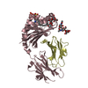
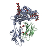
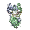
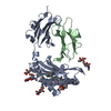
- PDB-5wcn: Structure of a bacterial polysialyltransferase in complex with CDP -
+ Open data

- Basic information
| Entry | Database: PDB / ID: 5wcn | |||||||||
|---|---|---|---|---|---|---|---|---|---|---|
| Title | Structure of a bacterial polysialyltransferase in complex with CDP | |||||||||
 Components Components | SiaD | |||||||||
 Keywords Keywords |  MEMBRANE PROTEIN / polysialyltransferase / GT-B MEMBRANE PROTEIN / polysialyltransferase / GT-B | |||||||||
| Function / homology | Alpha-2,8-polysialyltransferase / Alpha-2,8-polysialyltransferase (POLYST) / CYTIDINE-5'-DIPHOSPHATE / SiaD Function and homology information Function and homology information | |||||||||
| Biological species |  Mannheimia haemolytica (bacteria) Mannheimia haemolytica (bacteria) | |||||||||
| Method | X-RAY DIFFRACTION / SYNCHROTRON / MOLECULAR REPLACEMENT / Resolution: 3 Å | |||||||||
 Authors Authors | Worrall, L.J. / Lizak, C. / Strynadka, N.C.J. | |||||||||
| Funding support |  Canada, Canada,  United States, 2items United States, 2items
| |||||||||
 Citation Citation | Journal: Sci Rep / Year: 2017 Title: X-ray crystallographic structure of a bacterial polysialyltransferase provides insight into the biosynthesis of capsular polysialic acid. Authors: Lizak, C. / Worrall, L.J. / Baumann, L. / Pfleiderer, M.M. / Volkers, G. / Sun, T. / Sim, L. / Wakarchuk, W. / Withers, S.G. / Strynadka, N.C.J. | |||||||||
| History |
|
- Structure visualization
| Structure viewer | Molecule: MolmilJmol/JSmol |
|---|
- Downloads & links
-Download
| PDBx/mmCIF format | 5wcn.cif.gz | 170.3 KB | Display | PDBx/mmCIF format |
|---|---|---|---|---|
| PDB format | pdb5wcn.ent.gz | 134.8 KB | Display | PDB format |
| PDBx/mmJSON format | 5wcn.json.gz | Tree view | PDBx/mmJSON format | |
| Others |  Other downloads Other downloads |
-Validation report
| Arichive directory | https://data.pdbj.org/pub/pdb/validation_reports/wc/5wcnftp://data.pdbj.org/pub/pdb/validation_reports/wc/5wcn | HTTPS FTP |
|---|
-Related structure data


-Links
 PDBj
PDBj- Assembly
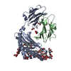
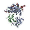
| Deposited unit | 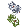
| ||||||||||||||||||
|---|---|---|---|---|---|---|---|---|---|---|---|---|---|---|---|---|---|---|---|
| 1 | 
| ||||||||||||||||||
| 2 | 
| ||||||||||||||||||
| Unit cell |
| ||||||||||||||||||
| Noncrystallographic symmetry (NCS) | NCS domain:
NCS domain segments: Component-ID: 0 / Ens-ID: 1 / Beg auth comp-ID: MET / Beg label comp-ID: MET / End auth comp-ID: ILE / End label comp-ID: ILE / Refine code: 0 / Auth seq-ID: 20 - 401 / Label seq-ID: 1 - 382
|
-Components
| #1: Protein | Mass: 45365.168 Da / Num. of mol.: 2 Source method: isolated from a genetically manipulated source Source: (gene. exp.) Mannheimia haemolytica (bacteria) / Gene: siaD / Production host: Escherichia coli (E. coli) / References: UniProt: G4RIN4#2: Chemical | Sulfate  Mass: 96.063 Da / Num. of mol.: 2 / Source method: obtained synthetically / Formula: SO4 Mass: 96.063 Da / Num. of mol.: 2 / Source method: obtained synthetically / Formula: SO4#3: Chemical | Cytidine diphosphate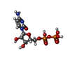  Mass: 403.176 Da / Num. of mol.: 2 / Source method: obtained synthetically / Formula: C9H15N3O11P2 Mass: 403.176 Da / Num. of mol.: 2 / Source method: obtained synthetically / Formula: C9H15N3O11P2#4: Water | ChemComp-HOH / | Water Mass: 18.015 Da / Num. of mol.: 32 / Source method: isolated from a natural source / Formula: H2O Mass: 18.015 Da / Num. of mol.: 32 / Source method: isolated from a natural source / Formula: H2O |
|---|
-Experimental details
-Experiment
| Experiment | Method: X-RAY DIFFRACTION / Number of used crystals: 1 |
|---|
- Sample preparation
| Crystal | Density Matthews: 2.96 Å3/Da / Density % sol: 58.43 % |
|---|---|
| Crystal grow | Temperature: 296 K / Method: vapor diffusion, sitting drop Details: 17% - 24% PEG3350 (v/v), 140 - 250 mM Mg2SO4 and 100 mM MES pH 7.2 |
-Data collection
| Diffraction | Mean temperature: 100 K | |||||||||||||||||||||
|---|---|---|---|---|---|---|---|---|---|---|---|---|---|---|---|---|---|---|---|---|---|---|
| Diffraction source | Source: SYNCHROTRON / Site: CLSI / Beamline: 08B1-1 / Wavelength: 1.07253 Å | |||||||||||||||||||||
| Detector | Type: RAYONIX MX300HE / Detector: CCD / Date: Dec 16, 2015 | |||||||||||||||||||||
| Radiation | Protocol: SINGLE WAVELENGTH / Monochromatic (M) / Laue (L): M / Scattering type: x-ray | |||||||||||||||||||||
| Radiation wavelength | Wavelength: 1.07253 Å / Relative weight: 1 | |||||||||||||||||||||
| Reflection | Resolution: 3→45.23 Å / Num. obs: 22618 / % possible obs: 99.9 % / Redundancy: 5.2 % / CC1/2: 0.996 / Rmerge(I) obs: 0.139 / Rpim(I) all: 0.067 / Rrim(I) all: 0.155 / Net I/σ(I): 8.8 / Num. measured all: 118669 / Scaling rejects: 0 | |||||||||||||||||||||
| Reflection shell | Diffraction-ID: 1
|
- Processing
| Software |
| ||||||||||||||||||||||||||||||||||||||||||||||||||||||||||||||||||||||||||||||||||||||||||||||||||||||||||||||||||||||||||||||||||||||||||||||||||||||||||||||||||||||||||||||||||||||
|---|---|---|---|---|---|---|---|---|---|---|---|---|---|---|---|---|---|---|---|---|---|---|---|---|---|---|---|---|---|---|---|---|---|---|---|---|---|---|---|---|---|---|---|---|---|---|---|---|---|---|---|---|---|---|---|---|---|---|---|---|---|---|---|---|---|---|---|---|---|---|---|---|---|---|---|---|---|---|---|---|---|---|---|---|---|---|---|---|---|---|---|---|---|---|---|---|---|---|---|---|---|---|---|---|---|---|---|---|---|---|---|---|---|---|---|---|---|---|---|---|---|---|---|---|---|---|---|---|---|---|---|---|---|---|---|---|---|---|---|---|---|---|---|---|---|---|---|---|---|---|---|---|---|---|---|---|---|---|---|---|---|---|---|---|---|---|---|---|---|---|---|---|---|---|---|---|---|---|---|---|---|---|---|
| Refinement | Method to determine structure: MOLECULAR REPLACEMENT / Resolution: 3→45.23 Å / Cor.coef. Fo:Fc: 0.949 / Cor.coef. Fo:Fc free: 0.912 / SU B: 20.039 / SU ML: 0.346 / Cross valid method: THROUGHOUT / ESU R Free: 0.426 / Stereochemistry target values: MAXIMUM LIKELIHOOD / Details: HYDROGENS HAVE BEEN ADDED IN THE RIDING POSITIONS
| ||||||||||||||||||||||||||||||||||||||||||||||||||||||||||||||||||||||||||||||||||||||||||||||||||||||||||||||||||||||||||||||||||||||||||||||||||||||||||||||||||||||||||||||||||||||
| Solvent computation | Ion probe radii: 0.8 Å / Shrinkage radii: 0.8 Å / VDW probe radii: 1.2 Å / Solvent model: MASK | ||||||||||||||||||||||||||||||||||||||||||||||||||||||||||||||||||||||||||||||||||||||||||||||||||||||||||||||||||||||||||||||||||||||||||||||||||||||||||||||||||||||||||||||||||||||
| Displacement parameters | Biso mean: 81.699 Å2
| ||||||||||||||||||||||||||||||||||||||||||||||||||||||||||||||||||||||||||||||||||||||||||||||||||||||||||||||||||||||||||||||||||||||||||||||||||||||||||||||||||||||||||||||||||||||
| Refinement step | Cycle: 1 / Resolution: 3→45.23 Å
| ||||||||||||||||||||||||||||||||||||||||||||||||||||||||||||||||||||||||||||||||||||||||||||||||||||||||||||||||||||||||||||||||||||||||||||||||||||||||||||||||||||||||||||||||||||||
| Refine LS restraints |
|