[English] 日本語
Yorodumi
- PDB-5w6q: Structural basis for recognition of artificial DNA by an evolved ... -

+
Open data


ID or keywords:

Loading...

-
Basic information

Entry
Database: PDB / ID: 5w6q
Title
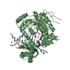
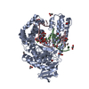
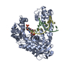
Structural basis for recognition of artificial DNA by an evolved KlenTaq variant
Components
  • DNA (5'-D(*GP*AP*CP*CP*AP*CP*GP*GP*CP*GP*CP*(1W5))-3')
  • DNA (5'-D(P*GP*(1WA)P*GP*CP*GP*CP*CP*GP*TP*GP*GP*TP*C)-3')
  • DNA polymerase I, thermostable
KeywordsDNA BINDING PROTEIN/DNA / Protein-DNA / AEGIS / unnatural base pair / host-guest system / transferase-dna complex / DNA BINDING PROTEIN / DNA BINDING PROTEIN-DNA complex
Function / homology
Function and homology information


nucleoside binding / double-strand break repair via alternative nonhomologous end joining / hydrolase activity, acting on ester bonds / DNA-templated DNA replication / DNA-directed DNA polymerase / DNA-directed DNA polymerase activity / DNA binding
Similarity search - Function
Taq polymerase, thermostable, exonuclease region / Taq polymerase, exonuclease / DNA polymerase I-like, H3TH domain / 5'-3' exonuclease, C-terminal SAM fold / 5'-3' exonuclease, alpha-helical arch, N-terminal / 5'-3' exonuclease, N-terminal resolvase-like domain / 5'-3' exonuclease / 5'-3' exonuclease / Taq DNA Polymerase; Chain T, domain 4 / Taq DNA Polymerase; Chain T, domain 4 ...Taq polymerase, thermostable, exonuclease region / Taq polymerase, exonuclease / DNA polymerase I-like, H3TH domain / 5'-3' exonuclease, C-terminal SAM fold / 5'-3' exonuclease, alpha-helical arch, N-terminal / 5'-3' exonuclease, N-terminal resolvase-like domain / 5'-3' exonuclease / 5'-3' exonuclease / Taq DNA Polymerase; Chain T, domain 4 / Taq DNA Polymerase; Chain T, domain 4 / DNA polymerase 1 / Alpha-Beta Plaits - #370 / Helix-hairpin-helix motif, class 2 / Helix-hairpin-helix class 2 (Pol1 family) motifs / 5'-3' exonuclease, C-terminal domain superfamily / DNA polymerase A / DNA polymerase family A / DNA-directed DNA polymerase, family A, conserved site / DNA polymerase family A signature. / DNA-directed DNA polymerase, family A, palm domain / DNA polymerase A domain / PIN-like domain superfamily / Helix-hairpin-helix DNA-binding motif, class 1 / Helix-hairpin-helix DNA-binding motif class 1 / Ribonuclease H-like superfamily/Ribonuclease H / Nucleotidyltransferase; domain 5 / Ribonuclease H superfamily / Ribonuclease H-like superfamily / Alpha-Beta Plaits / DNA/RNA polymerase superfamily / Up-down Bundle / 2-Layer Sandwich / Mainly Alpha / Alpha Beta
Similarity search - Domain/homology
DNA / DNA (> 10) / DNA polymerase I, thermostable
Similarity search - Component
Biological speciesThermus aquaticus (bacteria)
Escherichia coli (E. coli)
MethodX-RAY DIFFRACTION / SYNCHROTRON / MOLECULAR REPLACEMENT / molecular replacement / Resolution: 2.66 Å
AuthorsSingh, I. / Georgiadis, M.M.
CitationJournal: Nucleic Acids Res. / Year: 2018
Title: Snapshots of an evolved DNA polymerase pre- and post-incorporation of an unnatural nucleotide.
Authors: Singh, I. / Laos, R. / Hoshika, S. / Benner, S.A. / Georgiadis, M.M.
History
DepositionJun 16, 2017Deposition site: RCSB / Processing site: RCSB
Revision 1.0Jul 18, 2018Provider: repository / Type: Initial release
Revision 1.1Sep 19, 2018Group: Data collection / Database references / Category: citation
Item: _citation.journal_volume / _citation.page_first / _citation.page_last
Revision 1.2Oct 4, 2023Group: Data collection / Database references ...Data collection / Database references / Derived calculations / Refinement description
Category: chem_comp_atom / chem_comp_bond ...chem_comp_atom / chem_comp_bond / database_2 / pdbx_initial_refinement_model / struct_conn
Item: _database_2.pdbx_DOI / _database_2.pdbx_database_accession / _struct_conn.pdbx_leaving_atom_flag

-
Structure visualization

Structure viewerMolecule:
MolmilJmol/JSmol

Downloads & links

-
Assembly

Deposited unit
A: DNA polymerase I, thermostable
B: DNA (5'-D(*GP*AP*CP*CP*AP*CP*GP*GP*CP*GP*CP*(1W5))-3')
D: DNA (5'-D(P*GP*(1WA)P*GP*CP*GP*CP*CP*GP*TP*GP*GP*TP*C)-3')
C: DNA polymerase I, thermostable
E: DNA (5'-D(*GP*AP*CP*CP*AP*CP*GP*GP*CP*GP*CP*(1W5))-3')
F: DNA (5'-D(P*GP*(1WA)P*GP*CP*GP*CP*CP*GP*TP*GP*GP*TP*C)-3')
G: DNA polymerase I, thermostable
H: DNA (5'-D(*GP*AP*CP*CP*AP*CP*GP*GP*CP*GP*CP*(1W5))-3')
I: DNA (5'-D(P*GP*(1WA)P*GP*CP*GP*CP*CP*GP*TP*GP*GP*TP*C)-3')


Theoretical massNumber of molelcules
Total (without water)205,7019
Polymers205,7019
Non-polymers00
Water5,314295
1
A: DNA polymerase I, thermostable
B: DNA (5'-D(*GP*AP*CP*CP*AP*CP*GP*GP*CP*GP*CP*(1W5))-3')
D: DNA (5'-D(P*GP*(1WA)P*GP*CP*GP*CP*CP*GP*TP*GP*GP*TP*C)-3')


Theoretical massNumber of molelcules
Total (without water)68,5673
Polymers68,5673
Non-polymers00
Water543
TypeNameSymmetry operationNumber
identity operation1_555x,y,z1
Buried area4730 Å2
ΔGint-17 kcal/mol
Surface area24830 Å2
MethodPISA
2
C: DNA polymerase I, thermostable
E: DNA (5'-D(*GP*AP*CP*CP*AP*CP*GP*GP*CP*GP*CP*(1W5))-3')
F: DNA (5'-D(P*GP*(1WA)P*GP*CP*GP*CP*CP*GP*TP*GP*GP*TP*C)-3')


Theoretical massNumber of molelcules
Total (without water)68,5673
Polymers68,5673
Non-polymers00
Water543
TypeNameSymmetry operationNumber
identity operation1_555x,y,z1
Buried area4770 Å2
ΔGint-19 kcal/mol
Surface area24890 Å2
MethodPISA
3
G: DNA polymerase I, thermostable
H: DNA (5'-D(*GP*AP*CP*CP*AP*CP*GP*GP*CP*GP*CP*(1W5))-3')
I: DNA (5'-D(P*GP*(1WA)P*GP*CP*GP*CP*CP*GP*TP*GP*GP*TP*C)-3')


Theoretical massNumber of molelcules
Total (without water)68,5673
Polymers68,5673
Non-polymers00
Water543
TypeNameSymmetry operationNumber
identity operation1_555x,y,z1
Buried area4650 Å2
ΔGint-18 kcal/mol
Surface area25100 Å2
MethodPISA
Unit cell
Length a, b, c (Å)199.282, 114.611, 90.516
Angle α, β, γ (deg.)90.000, 91.880, 90.000
Int Tables number5
Space group name H-MC121
Noncrystallographic symmetry (NCS)NCS domain:
IDEns-IDDetails
11chain D and segid D
21chain F and segid F
31chain I and segid I

NCS domain segments:
Dom-IDComponent-IDEns-IDSelection detailsAuth asym-IDAuth seq-ID
111chain D and segid DD0
211chain F and segid FF0
311chain I and segid II0

-
Components

#1: Protein DNA polymerase I, thermostable / / Taq polymerase 1


Mass: 60862.902 Da / Num. of mol.: 3
Source method: isolated from a genetically manipulated source
Source: (gene. exp.) Thermus aquaticus (bacteria) / Gene: polA, pol1 / Production host: Escherichia coli (E. coli) / References: UniProt: P19821, DNA-directed DNA polymerase
#2: DNA chain DNA (5'-D(*GP*AP*CP*CP*AP*CP*GP*GP*CP*GP*CP*(1W5))-3')


Mass: 3677.380 Da / Num. of mol.: 3 / Source method: obtained synthetically / Source: (synth.) Escherichia coli (E. coli)
#3: DNA chain DNA (5'-D(P*GP*(1WA)P*GP*CP*GP*CP*CP*GP*TP*GP*GP*TP*C)-3')


Mass: 4026.612 Da / Num. of mol.: 3 / Source method: obtained synthetically / Source: (synth.) Escherichia coli (E. coli)
#4: Water ChemComp-HOH / water / Water


Mass: 18.015 Da / Num. of mol.: 295 / Source method: isolated from a natural source / Formula: H2O

-
Experimental details

-
Experiment

ExperimentMethod: X-RAY DIFFRACTION / Number of used crystals: 1

-
Sample preparation

CrystalDensity Matthews: 2.51 Å3/Da / Density % sol: 51.02 %
Crystal growTemperature: 277.15 K / Method: vapor diffusion, hanging drop / pH: 6 / Details: 14 % PEG 8000, 0.2 M Mg formate, 0.1 M MES pH 6.0

-
Data collection

DiffractionMean temperature: 100 K
Diffraction sourceSource: SYNCHROTRON / Site: APS / Beamline: 31-ID / Wavelength: 0.9793 Å
DetectorType: RAYONIX MX-225 / Detector: CCD / Date: Feb 24, 2016
RadiationProtocol: SINGLE WAVELENGTH / Monochromatic (M) / Laue (L): M / Scattering type: x-ray
Radiation wavelengthWavelength: 0.9793 Å / Relative weight: 1
ReflectionResolution: 2.662→49.132 Å / Num. all: 57514 / Num. obs: 57623 / % possible obs: 98.8 % / Redundancy: 3.4 % / Biso Wilson estimate: 39.82 Å2 / Rpim(I) all: 0.063 / Rrim(I) all: 0.118 / Rsym value: 0.082 / Net I/av σ(I): 8.9 / Net I/σ(I): 11.8 / Num. measured all: 194839
Reflection shell

Diffraction-ID: 1

Resolution (Å)Redundancy (%)Rmerge(I) obsMean I/σ(I) obsNum. measured allNum. unique allRpim(I) allRrim(I) allRsym valueNet I/σ(I) obs% possible all
2.66-2.812.70.4521.72212182980.3490.5990.4522.297.8
2.81-2.9830.3082.42357179050.2350.4180.3083.398.8
2.98-3.183.30.23.72447874620.1470.2710.25.299.2
3.18-3.443.50.12562465169710.0890.1690.1258.599.4
3.44-3.763.70.098.32365864230.0620.120.0912.699.1
3.76-4.213.80.06112.22178457880.0420.0810.06116.999.1
4.21-4.863.70.04715.61900550980.0320.0630.04721.998.5
4.86-5.953.70.04515.21626843530.0310.0610.04522.399
5.95-8.423.70.03420.41261033700.0240.0460.03425.799.1
8.42-99.3393.60.02129.2669318460.0150.0280.02135.496.8

-
Phasing

PhasingMethod: molecular replacement

-
Processing

Software
NameVersionClassification
PHENIX1.9_1692refinement
SCALAdata scaling
XDSdata collection
PDB_EXTRACT3.22data extraction
MOSFLMdata reduction
MOLREPphasing
RefinementMethod to determine structure: MOLECULAR REPLACEMENT
Starting model: 3SZ2
Resolution: 2.66→49.132 Å / SU ML: 0.36 / Cross valid method: FREE R-VALUE / σ(F): 1.35 / Phase error: 27.29 / Stereochemistry target values: ML
RfactorNum. reflection% reflection
Rfree0.2471 2818 4.89 %
Rwork0.2113 54805 -
obs0.2131 57623 98.53 %
Solvent computationShrinkage radii: 0.9 Å / VDW probe radii: 1.11 Å / Solvent model: FLAT BULK SOLVENT MODEL
Displacement parametersBiso max: 89.05 Å2 / Biso mean: 39.9312 Å2 / Biso min: 17.79 Å2
Refinement stepCycle: final / Resolution: 2.66→49.132 Å
ProteinNucleic acidLigandSolventTotal
Num. atoms12321 1542 0 295 14158
Biso mean---35.03 -
Num. residues----1638
Refine LS restraints
Refine-IDTypeDev idealNumber
X-RAY DIFFRACTIONf_bond_d0.00214329
X-RAY DIFFRACTIONf_angle_d0.70319733
X-RAY DIFFRACTIONf_chiral_restr0.0492193
X-RAY DIFFRACTIONf_plane_restr0.0032308
X-RAY DIFFRACTIONf_dihedral_angle_d15.4225486
Refine LS restraints NCS
Ens-IDDom-IDAuth asym-IDNumberRefine-IDRmsType
11D342X-RAY DIFFRACTION0.394TORSIONAL
12F342X-RAY DIFFRACTION0.394TORSIONAL
13I342X-RAY DIFFRACTION0.394TORSIONAL
LS refinement shell

Refine-ID: X-RAY DIFFRACTION / Rfactor Rfree error: 0 / Total num. of bins used: 20

Resolution (Å)Rfactor RfreeNum. reflection RfreeRfactor RworkNum. reflection RworkNum. reflection all% reflection obs (%)
2.66-2.70590.36911440.29782663280797
2.7059-2.75510.33061210.28222749287098
2.7551-2.80810.32481470.27522710285798
2.8081-2.86540.31111310.27732747287898
2.8654-2.92770.29891230.2762734285799
2.9277-2.99580.36461280.27512702283099
2.9958-3.07070.36141220.2762782290499
3.0707-3.15370.28471130.25932790290399
3.1537-3.24650.27951470.25392753290099
3.2465-3.35120.3051660.23432728289499
3.3512-3.4710.29051260.21662772289899
3.471-3.60990.24341510.21482736288799
3.6099-3.77410.22041240.19862729285399
3.7741-3.9730.24471400.18852766290699
3.973-4.22180.20711750.17322713288899
4.2218-4.54760.19521160.16552755287198
4.5476-5.00480.22021570.17212747290498
5.0048-5.72810.22781550.17992754290999
5.7281-7.21320.21321640.19222748291299
7.2132-49.14010.18241680.18072727289596

+
About Yorodumi

-
News

-
Feb 9, 2022. New format data for meta-information of EMDB entries

New format data for meta-information of EMDB entries

  • Version 3 of the EMDB header file is now the official format.
  • The previous official version 1.9 will be removed from the archive.

Related info.:EMDB header

External links:wwPDB to switch to version 3 of the EMDB data model

-
Aug 12, 2020. Covid-19 info

Covid-19 info

URL: https://pdbj.org/emnavi/covid19.php

New page: Covid-19 featured information page in EM Navigator.

Related info.:Covid-19 info / Mar 5, 2020. Novel coronavirus structure data

+
Mar 5, 2020. Novel coronavirus structure data

Novel coronavirus structure data

Related info.:Yorodumi Speices / Aug 12, 2020. Covid-19 info

External links:COVID-19 featured content - PDBj / Molecule of the Month (242):Coronavirus Proteases

+
Jan 31, 2019. EMDB accession codes are about to change! (news from PDBe EMDB page)

EMDB accession codes are about to change! (news from PDBe EMDB page)

  • The allocation of 4 digits for EMDB accession codes will soon come to an end. Whilst these codes will remain in use, new EMDB accession codes will include an additional digit and will expand incrementally as the available range of codes is exhausted. The current 4-digit format prefixed with “EMD-” (i.e. EMD-XXXX) will advance to a 5-digit format (i.e. EMD-XXXXX), and so on. It is currently estimated that the 4-digit codes will be depleted around Spring 2019, at which point the 5-digit format will come into force.
  • The EM Navigator/Yorodumi systems omit the EMD- prefix.

Related info.:Q: What is EMD? / ID/Accession-code notation in Yorodumi/EM Navigator

External links:EMDB Accession Codes are Changing Soon! / Contact to PDBj

+
Jul 12, 2017. Major update of PDB

Major update of PDB

  • wwPDB released updated PDB data conforming to the new PDBx/mmCIF dictionary.
  • This is a major update changing the version number from 4 to 5, and with Remediation, in which all the entries are updated.
  • In this update, many items about electron microscopy experimental information are reorganized (e.g. em_software).
  • Now, EM Navigator and Yorodumi are based on the updated data.

External links:wwPDB Remediation / Enriched Model Files Conforming to OneDep Data Standards Now Available in the PDB FTP Archive

-
Yorodumi

Thousand views of thousand structures

  • Yorodumi is a browser for structure data from EMDB, PDB, SASBDB, etc.
  • This page is also the successor to EM Navigator detail page, and also detail information page/front-end page for Omokage search.
  • The word "yorodu" (or yorozu) is an old Japanese word meaning "ten thousand". "mi" (miru) is to see.

Related info.:EMDB / PDB / SASBDB / Comparison of 3 databanks / Yorodumi Search / Aug 31, 2016. New EM Navigator & Yorodumi / Yorodumi Papers / Jmol/JSmol / Function and homology information / Changes in new EM Navigator and Yorodumi

Read more