 Movie
Movie Controller


+ Open data

- Basic information
| Entry | Database: PDB / ID: 5w0t | ||||||||||||||||||||||||
|---|---|---|---|---|---|---|---|---|---|---|---|---|---|---|---|---|---|---|---|---|---|---|---|---|---|
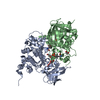
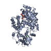
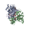
| Title | Crystal structure of monomeric Msp1 from S. cerevisiae | ||||||||||||||||||||||||
 Components Components | Protein MSP1 | ||||||||||||||||||||||||
 Keywords Keywords |  HYDROLASE / AAA ATPase HYDROLASE / AAA ATPase | ||||||||||||||||||||||||
| Function / homology |  Function and homology information Function and homology informationextraction of mislocalized protein from mitochondrial outer membrane / Class I peroxisomal membrane protein import / membrane protein dislocase activity / Translocases; Catalysing the translocation of amino acids and peptides; Linked to the hydrolysis of a nucleoside triphosphate / protein targeting to mitochondrion / protein hexamerization / peroxisomal membrane / mitochondrial outer membrane / membrane => GO:0016020 / ATP hydrolysis activity ...extraction of mislocalized protein from mitochondrial outer membrane / Class I peroxisomal membrane protein import / membrane protein dislocase activity / Translocases; Catalysing the translocation of amino acids and peptides; Linked to the hydrolysis of a nucleoside triphosphate / protein targeting to mitochondrion / protein hexamerization / peroxisomal membrane / mitochondrial outer membrane / membrane => GO:0016020 / ATP hydrolysis activity / mitochondrion / ATP bindingSimilarity search - Function | ||||||||||||||||||||||||
| Biological species |  Saccharomyces cerevisiae (brewer's yeast) Saccharomyces cerevisiae (brewer's yeast) | ||||||||||||||||||||||||
| Method | X-RAY DIFFRACTION / SYNCHROTRON / SAD / Resolution: 2.63 Å | ||||||||||||||||||||||||
 Authors Authors | Keenan, R.J. / Wohlever, M.L. / Mateja, A.M. | ||||||||||||||||||||||||
| Funding support |  United States, 7items United States, 7items
| ||||||||||||||||||||||||
 Citation Citation | Journal: Mol. Cell / Year: 2017 Title: Msp1 Is a Membrane Protein Dislocase for Tail-Anchored Proteins. Authors: Wohlever, M.L. / Mateja, A. / McGilvray, P.T. / Day, K.J. / Keenan, R.J. #1: Journal: EMBO J. / Year: 2014 Title: Msp1/ATAD1 maintains mitochondrial function by facilitating the degradation of mislocalized tail-anchored proteins. Authors: Chen, Y.C. / Umanah, G.K. / Dephoure, N. / Andrabi, S.A. / Gygi, S.P. / Dawson, T.M. / Dawson, V.L. / Rutter, J. #2: Journal: Proc. Natl. Acad. Sci. U.S.A. / Year: 2014 Title: The conserved AAA-ATPase Msp1 confers organelle specificity to tail-anchored proteins. Authors: Okreglak, V. / Walter, P. #3: Journal: Cell / Year: 2011 Title: The AAA+ ATPase Thorase regulates AMPA receptor-dependent synaptic plasticity and behavior. Authors: Zhang, J. / Wang, Y. / Chi, Z. / Keuss, M.J. / Pai, Y.M. / Kang, H.C. / Shin, J.H. / Bugayenko, A. / Wang, H. / Xiong, Y. / Pletnikov, M.V. / Mattson, M.P. / Dawson, T.M. / Dawson, V.L. | ||||||||||||||||||||||||
| History |
|
- Structure visualization
| Structure viewer | Molecule: MolmilJmol/JSmol |
|---|
- Downloads & links
-Download
| PDBx/mmCIF format | 5w0t.cif.gz | 133.4 KB | Display | PDBx/mmCIF format |
|---|---|---|---|---|
| PDB format | pdb5w0t.ent.gz | 109.5 KB | Display | PDB format |
| PDBx/mmJSON format | 5w0t.json.gz | Tree view | PDBx/mmJSON format | |
| Others |  Other downloads Other downloads |
-Validation report
| Arichive directory | https://data.pdbj.org/pub/pdb/validation_reports/w0/5w0tftp://data.pdbj.org/pub/pdb/validation_reports/w0/5w0t | HTTPS FTP |
|---|
-Related structure data
| Similar structure data |
|---|


-Links
 PDBj
PDBj


- Assembly
| Deposited unit | 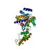
| ||||||||
|---|---|---|---|---|---|---|---|---|---|
| 1 | 
| ||||||||
| Unit cell |
|
-Components
| #1: Protein | Mass: 34343.125 Da / Num. of mol.: 1 / Fragment: UNP residues 51-345 Source method: isolated from a genetically manipulated source Source: (gene. exp.) Saccharomyces cerevisiae (brewer's yeast)Strain: ATCC 204508 / S288c / Gene: MSP1, YTA4, YGR028W / Production host:  Escherichia coli (E. coli) / Strain (production host): BL21(DE3)/pRIL / References: UniProt: P28737 Escherichia coli (E. coli) / Strain (production host): BL21(DE3)/pRIL / References: UniProt: P28737 | ||
|---|---|---|---|
| #2: Chemical | ChemComp-EDO / Ethylene glycol  Mass: 62.068 Da / Num. of mol.: 5 / Source method: obtained synthetically / Formula: C2H6O2 Mass: 62.068 Da / Num. of mol.: 5 / Source method: obtained synthetically / Formula: C2H6O2#3: Water | ChemComp-HOH / | Water Mass: 18.015 Da / Num. of mol.: 11 / Source method: isolated from a natural source / Formula: H2O Mass: 18.015 Da / Num. of mol.: 11 / Source method: isolated from a natural source / Formula: H2O |
-Experimental details
-Experiment
| Experiment | Method: X-RAY DIFFRACTION / Number of used crystals: 1 |
|---|
- Sample preparation
| Crystal | Density Matthews: 2.76 Å3/Da / Density % sol: 55.41 % |
|---|---|
| Crystal grow | Temperature: 293 K / Method: vapor diffusion, hanging drop Details: ~9 mg/ml protein in 20 mM Hepes pH 7.5, 100 mM NaCl and 1 mM DTT was mixed with reservoir solution containing 16% PEG3350 and 0.6 M Sodium Thiocyanate |
-Data collection
| Diffraction | Mean temperature: 100 K |
|---|---|
| Diffraction source | Source: SYNCHROTRON / Site: APS / Beamline: 24-ID-C / Wavelength: 0.97918 Å |
| Detector | Type: PSI PILATUS 6M / Detector: PIXEL / Date: Oct 30, 2015 |
| Radiation | Protocol: SINGLE WAVELENGTH / Monochromatic (M) / Laue (L): M / Scattering type: x-ray |
| Radiation wavelength | Wavelength: 0.97918 Å / Relative weight: 1 |
| Reflection | Resolution: 2.63→206.71 Å / Num. obs: 11608 / % possible obs: 100 % / Redundancy: 17.4 % / CC1/2: 0.99 / Rmerge(I) obs: 0.211 / Rpim(I) all: 0.051 / Net I/σ(I): 9.9 |
| Reflection shell | Resolution: 2.63→2.7 Å / Redundancy: 9.6 % / Rmerge(I) obs: 1.447 / Mean I/σ(I) obs: 1.2 / Num. unique obs: 843 / CC1/2: 0.762 / Rpim(I) all: 0.491 / % possible all: 100 |
- Processing
| Software |
| ||||||||||||||||||||||||||||||||||||||||||||||||||||||||||||||||||||||||||||||||||||||||||||||||||||
|---|---|---|---|---|---|---|---|---|---|---|---|---|---|---|---|---|---|---|---|---|---|---|---|---|---|---|---|---|---|---|---|---|---|---|---|---|---|---|---|---|---|---|---|---|---|---|---|---|---|---|---|---|---|---|---|---|---|---|---|---|---|---|---|---|---|---|---|---|---|---|---|---|---|---|---|---|---|---|---|---|---|---|---|---|---|---|---|---|---|---|---|---|---|---|---|---|---|---|---|---|---|
| Refinement | Method to determine structure: SAD / Resolution: 2.63→68.902 Å / SU ML: 0.33 / Cross valid method: THROUGHOUT / σ(F): 1.33 / Phase error: 33.02
| ||||||||||||||||||||||||||||||||||||||||||||||||||||||||||||||||||||||||||||||||||||||||||||||||||||
| Solvent computation | Shrinkage radii: 0.9 Å / VDW probe radii: 1.11 Å | ||||||||||||||||||||||||||||||||||||||||||||||||||||||||||||||||||||||||||||||||||||||||||||||||||||
| Displacement parameters | Biso max: 261.93 Å2 / Biso mean: 76.4023 Å2 / Biso min: 40.07 Å2 | ||||||||||||||||||||||||||||||||||||||||||||||||||||||||||||||||||||||||||||||||||||||||||||||||||||
| Refinement step | Cycle: final / Resolution: 2.63→68.902 Å
| ||||||||||||||||||||||||||||||||||||||||||||||||||||||||||||||||||||||||||||||||||||||||||||||||||||
| Refine LS restraints |
| ||||||||||||||||||||||||||||||||||||||||||||||||||||||||||||||||||||||||||||||||||||||||||||||||||||
| LS refinement shell | Refine-ID: X-RAY DIFFRACTION / Rfactor Rfree error: 0 / Total num. of bins used: 4
| ||||||||||||||||||||||||||||||||||||||||||||||||||||||||||||||||||||||||||||||||||||||||||||||||||||
| Refinement TLS params. | Method: refined / Refine-ID: X-RAY DIFFRACTION
| ||||||||||||||||||||||||||||||||||||||||||||||||||||||||||||||||||||||||||||||||||||||||||||||||||||
| Refinement TLS group |
|
