 Movie
Movie Controller

[English] 日本語
 Yorodumi
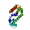
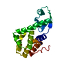
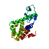
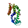
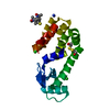
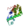
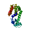
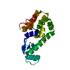
- PDB-5vnr: X-ray structure of perdeuterated T4 lysozyme cysteine-free pseudo... -
+ Open data

- Basic information
| Entry | Database: PDB / ID: 5vnr | ||||||
|---|---|---|---|---|---|---|---|
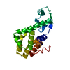
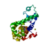
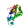
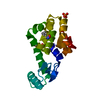
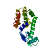
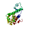
| Title | X-ray structure of perdeuterated T4 lysozyme cysteine-free pseudo-wild type at cryogenic temperature | ||||||
 Components Components | Endolysin Lysin Lysin | ||||||
 Keywords Keywords | HYDROLASE / T4 lysozyme / Neutron Crystallography / Hydrogen bonding network / Hydrogen bond / Water | ||||||
| Function / homology |  Function and homology information Function and homology informationviral release from host cell by cytolysis / peptidoglycan catabolic process / cell wall macromolecule catabolic process / lysozyme / lysozyme activity / host cell cytoplasm / defense response to bacteriumSimilarity search - Function | ||||||
| Biological species |  Enterobacteria phage T4 (virus) Enterobacteria phage T4 (virus) | ||||||
| Method | X-RAY DIFFRACTION / MOLECULAR REPLACEMENT / Resolution: 1.631 Å | ||||||
 Authors Authors | Li, L. / Shukla, S. / Meilleur, F. / Standaert, R.F. / Pierce, J. / Myles, D.A.A. / Cuneo, M.J. | ||||||
 Citation Citation | Journal: Protein Sci. / Year: 2017 Title: Neutron crystallographic studies of T4 lysozyme at cryogenic temperature. Authors: Li, L. / Shukla, S. / Meilleur, F. / Standaert, R.F. / Pierce, J. / Myles, D.A.A. / Cuneo, M.J. | ||||||
| History |
|
- Structure visualization
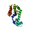
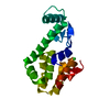
| Structure viewer | Molecule: MolmilJmol/JSmol |
|---|
- Downloads & links
-Download
| PDBx/mmCIF format | 5vnr.cif.gz | 57.9 KB | Display | PDBx/mmCIF format |
|---|---|---|---|---|
| PDB format | pdb5vnr.ent.gz | 39.6 KB | Display | PDB format |
| PDBx/mmJSON format | 5vnr.json.gz | Tree view | PDBx/mmJSON format | |
| Others |  Other downloads Other downloads |
-Validation report
| Arichive directory | https://data.pdbj.org/pub/pdb/validation_reports/vn/5vnrftp://data.pdbj.org/pub/pdb/validation_reports/vn/5vnr | HTTPS FTP |
|---|
-Related structure data
| Related structure data | 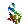 5vnqC 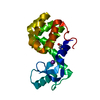 1lw9S C: citing same article ( S: Starting model for refinement |
|---|---|
| Similar structure data |


-Links
 PDBj
PDBj


- Assembly
| Deposited unit | 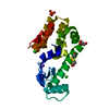
| ||||||||
|---|---|---|---|---|---|---|---|---|---|
| 1 |
| ||||||||
| Unit cell |
|
-Components
-Protein , 1 types, 1 molecules A
| #1: Protein | Lysin / Lysis protein / Lysozyme / Muramidase Mass: 18628.363 Da / Num. of mol.: 1 / Mutation: C54T, C97A Source method: isolated from a genetically manipulated source Source: (gene. exp.) Enterobacteria phage T4 (virus) / Gene: e, T4Tp126 / Production host:  Escherichia coli (E. coli) / References: UniProt: D9IEF7, UniProt: P00720*PLUS, lysozyme Escherichia coli (E. coli) / References: UniProt: D9IEF7, UniProt: P00720*PLUS, lysozyme |
|---|
-Non-polymers , 6 types, 299 molecules 


| #2: Chemical | ChemComp-PO4 / Phosphate Mass: 94.971 Da / Num. of mol.: 1 / Source method: obtained synthetically / Formula: PO4 Mass: 94.971 Da / Num. of mol.: 1 / Source method: obtained synthetically / Formula: PO4 | ||||
|---|---|---|---|---|---|
| #3: Chemical | ChemComp-NA /  Mass: 22.990 Da / Num. of mol.: 1 / Source method: obtained synthetically / Formula: Na Mass: 22.990 Da / Num. of mol.: 1 / Source method: obtained synthetically / Formula: Na | ||||
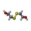
| #4: Chemical | ChemComp-HED /  Mass: 154.251 Da / Num. of mol.: 1 / Source method: obtained synthetically / Formula: C4H10O2S2 Mass: 154.251 Da / Num. of mol.: 1 / Source method: obtained synthetically / Formula: C4H10O2S2 | ||||
| #5: Chemical | Chloride Mass: 35.453 Da / Num. of mol.: 2 / Source method: obtained synthetically / Formula: Cl Mass: 35.453 Da / Num. of mol.: 2 / Source method: obtained synthetically / Formula: Cl#6: Chemical | ChemComp-GOL / | Glycerol Mass: 92.094 Da / Num. of mol.: 1 / Source method: obtained synthetically / Formula: C3H8O3 Mass: 92.094 Da / Num. of mol.: 1 / Source method: obtained synthetically / Formula: C3H8O3#7: Water | ChemComp-HOH / | WaterMass: 18.015 Da / Num. of mol.: 293 / Source method: isolated from a natural source / Formula: H2O |
-Experimental details
-Experiment
| Experiment | Method: X-RAY DIFFRACTION / Number of used crystals: 1 |
|---|
- Sample preparation
| Crystal | Density Matthews: 2.65 Å3/Da / Density % sol: 53.54 % |
|---|---|
| Crystal grow | Temperature: 293 K / Method: vapor diffusion, sitting drop Details: ~2.0 M Na/K phosphate, pH 6-7, 250 mM NaCl, 40mM 2-hydroxyethyl disulfide PH range: 6-7 |
-Data collection
| Diffraction | Mean temperature: 100 K |
|---|---|
| Diffraction source | Source: ROTATING ANODE / Type: RIGAKU MICROMAX-007 HF / Wavelength: 1.54 Å |
| Detector | Type: RIGAKU RAXIS IV++ / Detector: IMAGE PLATE / Date: Apr 4, 2017 |
| Radiation | Protocol: SINGLE WAVELENGTH / Monochromatic (M) / Laue (L): M / Scattering type: x-ray |
| Radiation wavelength | Wavelength: 1.54 Å / Relative weight: 1 |
| Reflection | Resolution: 1.63→50 Å / Num. obs: 25255 / % possible obs: 99.7 % / Redundancy: 10.1 % / Rmerge(I) obs: 0.096 / Net I/σ(I): 20.1 |
- Processing
| Software |
| |||||||||||||||||||||||||||||||||||||||||||||||||||||||||||||||||||||||||||||||||||||||||||||||||||||||||
|---|---|---|---|---|---|---|---|---|---|---|---|---|---|---|---|---|---|---|---|---|---|---|---|---|---|---|---|---|---|---|---|---|---|---|---|---|---|---|---|---|---|---|---|---|---|---|---|---|---|---|---|---|---|---|---|---|---|---|---|---|---|---|---|---|---|---|---|---|---|---|---|---|---|---|---|---|---|---|---|---|---|---|---|---|---|---|---|---|---|---|---|---|---|---|---|---|---|---|---|---|---|---|---|---|---|---|
| Refinement | Method to determine structure: MOLECULAR REPLACEMENT Starting model: 1LW9 Resolution: 1.631→28.546 Å / SU ML: 0.14 / Cross valid method: FREE R-VALUE / σ(F): 1.34 / Phase error: 15.73 / Stereochemistry target values: ML
| |||||||||||||||||||||||||||||||||||||||||||||||||||||||||||||||||||||||||||||||||||||||||||||||||||||||||
| Solvent computation | Shrinkage radii: 0.9 Å / VDW probe radii: 1.11 Å / Solvent model: FLAT BULK SOLVENT MODEL | |||||||||||||||||||||||||||||||||||||||||||||||||||||||||||||||||||||||||||||||||||||||||||||||||||||||||
| Refinement step | Cycle: LAST / Resolution: 1.631→28.546 Å
| |||||||||||||||||||||||||||||||||||||||||||||||||||||||||||||||||||||||||||||||||||||||||||||||||||||||||
| Refine LS restraints |
| |||||||||||||||||||||||||||||||||||||||||||||||||||||||||||||||||||||||||||||||||||||||||||||||||||||||||
| LS refinement shell |
|
