 Movie
Movie Controller


+ Open data

- Basic information
| Entry | Database: PDB / ID: 5vmn | ||||||
|---|---|---|---|---|---|---|---|
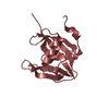
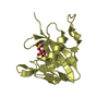
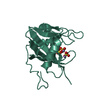
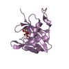
| Title | Crystal structure of grouper iridovirus GIV66 | ||||||
 Components Components | Bak protein | ||||||
 Keywords Keywords |  VIRAL PROTEIN / Apoptosis / Bcl-2 / iridovirus / grouper VIRAL PROTEIN / Apoptosis / Bcl-2 / iridovirus / grouper | ||||||
| Function / homology | : / Bcl-2, Bcl-2 homology region 1-3 / Bcl2-like / Apoptosis regulator proteins, Bcl-2 family / BCL2-like apoptosis inhibitors family profile. / Bcl-2-like superfamily / membrane / NITRATE ION / Bak protein Function and homology information Function and homology information | ||||||
| Biological species |  Grouper iridovirus Grouper iridovirus | ||||||
| Method | X-RAY DIFFRACTION / SYNCHROTRON / SAD / Resolution: 1.65 Å | ||||||
 Authors Authors | Banjara, S. / Kvansakul, M. | ||||||
 Citation Citation | Journal: J. Biol. Chem. / Year: 2018 Title: Grouper iridovirus GIV66 is a Bcl-2 protein that inhibits apoptosis by exclusively sequestering Bim. Authors: Banjara, S. / Mao, J. / Ryan, T.M. / Caria, S. / Kvansakul, M. | ||||||
| History |
|
- Structure visualization
| Structure viewer | Molecule: MolmilJmol/JSmol |
|---|
- Downloads & links
-Download
| PDBx/mmCIF format | 5vmn.cif.gz | 68.3 KB | Display | PDBx/mmCIF format |
|---|---|---|---|---|
| PDB format | pdb5vmn.ent.gz | 50.7 KB | Display | PDB format |
| PDBx/mmJSON format | 5vmn.json.gz | Tree view | PDBx/mmJSON format | |
| Others |  Other downloads Other downloads |
-Validation report
| Arichive directory | https://data.pdbj.org/pub/pdb/validation_reports/vm/5vmnftp://data.pdbj.org/pub/pdb/validation_reports/vm/5vmn | HTTPS FTP |
|---|
-Related structure data


-Links
 PDBj
PDBj
- Assembly
| Deposited unit | 
| ||||||||
|---|---|---|---|---|---|---|---|---|---|
| 1 |
| ||||||||
| Unit cell |
| ||||||||
| Components on special symmetry positions |
|
-Components
| #1: Protein | Mass: 14943.972 Da / Num. of mol.: 1 / Fragment: UNP residues 1-127 Source method: isolated from a genetically manipulated source Source: (gene. exp.) Grouper iridovirus / Gene: GIV66 / Plasmid: pGEX-6P3 / Production host:  Escherichia coli BL21(DE3) (bacteria) / References: UniProt: Q5GAF0 Escherichia coli BL21(DE3) (bacteria) / References: UniProt: Q5GAF0 | ||||
|---|---|---|---|---|---|
| #2: Chemical | ChemComp-NO3 / Nitrate  Mass: 62.005 Da / Num. of mol.: 8 / Source method: obtained synthetically / Formula: NO3 Mass: 62.005 Da / Num. of mol.: 8 / Source method: obtained synthetically / Formula: NO3#3: Chemical | ChemComp-EDO / Ethylene glycol  Mass: 62.068 Da / Num. of mol.: 6 / Source method: obtained synthetically / Formula: C2H6O2 Mass: 62.068 Da / Num. of mol.: 6 / Source method: obtained synthetically / Formula: C2H6O2#4: Water | ChemComp-HOH / | Water Mass: 18.015 Da / Num. of mol.: 106 / Source method: isolated from a natural source / Formula: H2O Mass: 18.015 Da / Num. of mol.: 106 / Source method: isolated from a natural source / Formula: H2O |
-Experimental details
-Experiment
| Experiment | Method: X-RAY DIFFRACTION / Number of used crystals: 1 |
|---|
- Sample preparation
| Crystal | Density Matthews: 3.32 Å3/Da / Density % sol: 63 % |
|---|---|
| Crystal grow | Temperature: 293 K / Method: vapor diffusion, sitting drop / pH: 4.6 Details: 4.0 M sodium nitrate with 0.1 M sodium acetate pH 4.6 |
-Data collection
| Diffraction | Mean temperature: 100 K |
|---|---|
| Diffraction source | Source: SYNCHROTRON / Site: Australian Synchrotron  / Beamline: MX1 / Wavelength: 0.9537 Å / Beamline: MX1 / Wavelength: 0.9537 Å |
| Detector | Type: ADSC QUANTUM 315r / Detector: CCD / Date: Nov 9, 2016 |
| Radiation | Protocol: SINGLE WAVELENGTH / Monochromatic (M) / Laue (L): M / Scattering type: x-ray |
| Radiation wavelength | Wavelength: 0.9537 Å / Relative weight: 1 |
| Reflection | Resolution: 1.65→42.91 Å / Num. obs: 24871 / % possible obs: 99.9 % / Redundancy: 10 % / CC1/2: 0.999 / Rmerge(I) obs: 0.057 / Net I/σ(I): 24.8 |
| Reflection shell | Resolution: 1.65→1.68 Å / Redundancy: 11.1 % / Rmerge(I) obs: 2.395 / Mean I/σ(I) obs: 1.1 / Num. unique obs: 1219 / CC1/2: 0.396 / % possible all: 100 |
- Processing
| Software |
| ||||||||||||||||||||||||||||||||||||||||||||||||||||||||||||||||||||||||||||||||||||||||||||||||||||
|---|---|---|---|---|---|---|---|---|---|---|---|---|---|---|---|---|---|---|---|---|---|---|---|---|---|---|---|---|---|---|---|---|---|---|---|---|---|---|---|---|---|---|---|---|---|---|---|---|---|---|---|---|---|---|---|---|---|---|---|---|---|---|---|---|---|---|---|---|---|---|---|---|---|---|---|---|---|---|---|---|---|---|---|---|---|---|---|---|---|---|---|---|---|---|---|---|---|---|---|---|---|
| Refinement | Method to determine structure: SAD / Resolution: 1.65→41.964 Å / SU ML: 0.24 / Cross valid method: FREE R-VALUE / σ(F): 1.33 / Phase error: 22.6 / Stereochemistry target values: ML
| ||||||||||||||||||||||||||||||||||||||||||||||||||||||||||||||||||||||||||||||||||||||||||||||||||||
| Solvent computation | Shrinkage radii: 0.9 Å / VDW probe radii: 1.11 Å / Solvent model: FLAT BULK SOLVENT MODEL | ||||||||||||||||||||||||||||||||||||||||||||||||||||||||||||||||||||||||||||||||||||||||||||||||||||
| Refinement step | Cycle: LAST / Resolution: 1.65→41.964 Å
| ||||||||||||||||||||||||||||||||||||||||||||||||||||||||||||||||||||||||||||||||||||||||||||||||||||
| Refine LS restraints |
| ||||||||||||||||||||||||||||||||||||||||||||||||||||||||||||||||||||||||||||||||||||||||||||||||||||
| LS refinement shell |
| ||||||||||||||||||||||||||||||||||||||||||||||||||||||||||||||||||||||||||||||||||||||||||||||||||||
| Refinement TLS params. | Method: refined / Refine-ID: X-RAY DIFFRACTION
| ||||||||||||||||||||||||||||||||||||||||||||||||||||||||||||||||||||||||||||||||||||||||||||||||||||
| Refinement TLS group |
|
