 Movie
Movie Controller

[English] 日本語
 Yorodumi
- PDB-5vj1: Crystal structure of a Pseudomonas malonate decarboxylase hetero-... -
+ Open data

- Basic information
| Entry | Database: PDB / ID: 5vj1 | ||||||
|---|---|---|---|---|---|---|---|
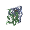
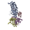
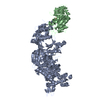
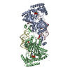
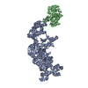
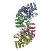
| Title | Crystal structure of a Pseudomonas malonate decarboxylase hetero-tetramer in complex with coenzyme A | ||||||
 Components Components |
| ||||||
 Keywords Keywords |  TRANSFERASE / acetyl-CoA carboxylase / coa transferase / acp transferase TRANSFERASE / acetyl-CoA carboxylase / coa transferase / acp transferase | ||||||
| Function / homology |  Function and homology informationmalonyl-S-ACP:biotin-protein carboxyltransferase / malonyl-CoA biosynthetic process / acetyl-CoA carboxylase activity / carboxy-lyase activity / acyl carrier activity / fatty acid biosynthetic process / transferase activity / carbohydrate metabolic process / cytoplasm Function and homology informationmalonyl-S-ACP:biotin-protein carboxyltransferase / malonyl-CoA biosynthetic process / acetyl-CoA carboxylase activity / carboxy-lyase activity / acyl carrier activity / fatty acid biosynthetic process / transferase activity / carbohydrate metabolic process / cytoplasmSimilarity search - Function | ||||||
| Biological species |   Pseudomonas aeruginosa (bacteria)Pseudomonas fluorescens (bacteria) Pseudomonas aeruginosa (bacteria)Pseudomonas fluorescens (bacteria) | ||||||
| Method | X-RAY DIFFRACTION / SYNCHROTRON / MOLECULAR REPLACEMENT / Resolution: 2.995 Å | ||||||
 Authors Authors | Maderbocus, R. / Tong, L. | ||||||
 Citation Citation | Journal: Nat Commun / Year: 2017 Title: Crystal structure of a Pseudomonas malonate decarboxylase holoenzyme hetero-tetramer. Authors: Maderbocus, R. / Fields, B.L. / Hamilton, K. / Luo, S. / Tran, T.H. / Dietrich, L.E.P. / Tong, L. | ||||||
| History |
|
- Structure visualization
| Structure viewer | Molecule: MolmilJmol/JSmol |
|---|
- Downloads & links
-Download
| PDBx/mmCIF format | 5vj1.cif.gz | 455.4 KB | Display | PDBx/mmCIF format |
|---|---|---|---|---|
| PDB format | pdb5vj1.ent.gz | 367.2 KB | Display | PDB format |
| PDBx/mmJSON format | 5vj1.json.gz | Tree view | PDBx/mmJSON format | |
| Others |  Other downloads Other downloads |
-Validation report
| Arichive directory | https://data.pdbj.org/pub/pdb/validation_reports/vj/5vj1ftp://data.pdbj.org/pub/pdb/validation_reports/vj/5vj1 | HTTPS FTP |
|---|
-Related structure data


-Links
 PDBj
PDBj


- Assembly
| Deposited unit | 
| ||||||||
|---|---|---|---|---|---|---|---|---|---|
| 1 | 
| ||||||||
| 2 | 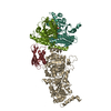
| ||||||||
| 3 | 
| ||||||||
| Unit cell |
|
-Components
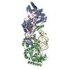
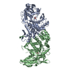
-Protein , 4 types, 8 molecules AICKDLEM
| #1: Protein | Mass: 61501.242 Da / Num. of mol.: 2 Source method: isolated from a genetically manipulated source Source: (gene. exp.) Pseudomonas aeruginosa (strain ATCC 15692 / DSM 22644 / CIP 104116 / JCM 14847 / LMG 12228 / 1C / PRS 101 / PAO1) (bacteria)Strain: ATCC 15692 / DSM 22644 / CIP 104116 / JCM 14847 / LMG 12228 / 1C / PRS 101 / PAO1 Gene: mdcA, PA0208 / Production host: Escherichia coli (E. coli) / References: UniProt: Q9I6T0#2: Protein | Mass: 10692.981 Da / Num. of mol.: 2 Source method: isolated from a genetically manipulated source Source: (gene. exp.) Pseudomonas fluorescens (strain ATCC BAA-477 / NRRL B-23932 / Pf-5) (bacteria)Strain: ATCC BAA-477 / NRRL B-23932 / Pf-5 / Gene: mdcC, PFL_5818 / Production host: Escherichia coli (E. coli) / References: UniProt: Q4K4F7#3: Protein | Mass: 30371.320 Da / Num. of mol.: 2 Source method: isolated from a genetically manipulated source Source: (gene. exp.) Pseudomonas aeruginosa (bacteria)Gene: madC, mdcD, AO964_31600, AOY09_06294, BH593_13640, PAERUG_E15_London_28_01_14_07061, PAERUG_P32_London_17_VIM_2_10_11_04127, PAMH19_0209 Production host: Escherichia coli (E. coli)References: UniProt: A0A071KS24, UniProt: Q9I6S7*PLUS, malonyl-S-ACP:biotin-protein carboxyltransferase, methylmalonyl-CoA carboxytransferase#4: Protein | Mass: 30351.605 Da / Num. of mol.: 2 Source method: isolated from a genetically manipulated source Source: (gene. exp.) Pseudomonas aeruginosa (bacteria)Gene: madD, AO964_31595, AOY09_06293, PAERUG_E15_London_28_01_14_07062, PAERUG_P32_London_17_VIM_2_10_11_04128 Production host: Escherichia coli (E. coli)References: UniProt: A0A0C6EV56, UniProt: Q9I6S6*PLUS, malonyl-S-ACP:biotin-protein carboxyltransferase |
|---|
-Non-polymers , 2 types, 3 molecules 

| #5: Chemical | ChemComp-CL / Chloride Mass: 35.453 Da / Num. of mol.: 1 / Source method: obtained synthetically / Formula: Cl Mass: 35.453 Da / Num. of mol.: 1 / Source method: obtained synthetically / Formula: Cl |
|---|---|
| #6: Chemical | Coenzyme A Mass: 767.534 Da / Num. of mol.: 2 / Source method: obtained synthetically / Formula: C21H36N7O16P3S Mass: 767.534 Da / Num. of mol.: 2 / Source method: obtained synthetically / Formula: C21H36N7O16P3S |
-Experimental details
-Experiment
| Experiment | Method: X-RAY DIFFRACTION / Number of used crystals: 1 |
|---|
- Sample preparation
| Crystal | Density Matthews: 3.15 Å3/Da / Density % sol: 60.91 % |
|---|---|
| Crystal grow | Temperature: 293 K / Method: vapor diffusion, sitting drop / pH: 8 / Details: 20% (w/v) PEG3350 and 8% Tacsimate / PH range: 8 |
-Data collection
| Diffraction | Mean temperature: 100 K |
|---|---|
| Diffraction source | Source: SYNCHROTRON / Site: APS  / Beamline: 24-ID-C / Wavelength: 0.9789 Å / Beamline: 24-ID-C / Wavelength: 0.9789 Å |
| Detector | Type: DECTRIS PILATUS 6M / Detector: PIXEL / Date: Apr 7, 2016 / Details: MIRRORS |
| Radiation | Monochromator: SI(111) / Protocol: SINGLE WAVELENGTH / Monochromatic (M) / Laue (L): M / Scattering type: x-ray |
| Radiation wavelength | Wavelength: 0.9789 Å / Relative weight: 1 |
| Reflection | Resolution: 3→50 Å / Num. obs: 63210 / % possible obs: 98.8 % / Observed criterion σ(I): -3 / Redundancy: 3.2 % / Rmerge(I) obs: 0.1 / Net I/σ(I): 12 |
| Reflection shell | Resolution: 3→3.18 Å / Redundancy: 3.3 % / Rmerge(I) obs: 0.679 / Mean I/σ(I) obs: 1.7 / % possible all: 97.9 |
- Processing
| Software |
| ||||||||||||||||||||||||||||||||||||||||||||||||||||||||||||||||||||||||||||||||||||||||||||||||||||||||||||||||||||||||||||||||||||||||||||||||||||||||||||||||||||||||
|---|---|---|---|---|---|---|---|---|---|---|---|---|---|---|---|---|---|---|---|---|---|---|---|---|---|---|---|---|---|---|---|---|---|---|---|---|---|---|---|---|---|---|---|---|---|---|---|---|---|---|---|---|---|---|---|---|---|---|---|---|---|---|---|---|---|---|---|---|---|---|---|---|---|---|---|---|---|---|---|---|---|---|---|---|---|---|---|---|---|---|---|---|---|---|---|---|---|---|---|---|---|---|---|---|---|---|---|---|---|---|---|---|---|---|---|---|---|---|---|---|---|---|---|---|---|---|---|---|---|---|---|---|---|---|---|---|---|---|---|---|---|---|---|---|---|---|---|---|---|---|---|---|---|---|---|---|---|---|---|---|---|---|---|---|---|---|---|---|---|
| Refinement | Method to determine structure: MOLECULAR REPLACEMENT / Resolution: 2.995→47.901 Å / SU ML: 0.42 / Cross valid method: THROUGHOUT / σ(F): 1.35 / Phase error: 26.79 / Stereochemistry target values: ML
| ||||||||||||||||||||||||||||||||||||||||||||||||||||||||||||||||||||||||||||||||||||||||||||||||||||||||||||||||||||||||||||||||||||||||||||||||||||||||||||||||||||||||
| Solvent computation | Shrinkage radii: 0.9 Å / VDW probe radii: 1.11 Å / Solvent model: FLAT BULK SOLVENT MODEL | ||||||||||||||||||||||||||||||||||||||||||||||||||||||||||||||||||||||||||||||||||||||||||||||||||||||||||||||||||||||||||||||||||||||||||||||||||||||||||||||||||||||||
| Refinement step | Cycle: LAST / Resolution: 2.995→47.901 Å
| ||||||||||||||||||||||||||||||||||||||||||||||||||||||||||||||||||||||||||||||||||||||||||||||||||||||||||||||||||||||||||||||||||||||||||||||||||||||||||||||||||||||||
| Refine LS restraints |
| ||||||||||||||||||||||||||||||||||||||||||||||||||||||||||||||||||||||||||||||||||||||||||||||||||||||||||||||||||||||||||||||||||||||||||||||||||||||||||||||||||||||||
| LS refinement shell |
|
