 Movie
Movie Controller

[English] 日本語
 Yorodumi
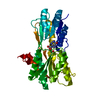
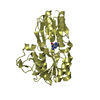
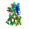
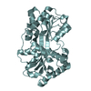
- PDB-5vik: Crystal structure of monomeric near-infrared fluorescent protein ... -
+ Open data

- Basic information
| Entry | Database: PDB / ID: 5vik | ||||||
|---|---|---|---|---|---|---|---|
| Title | Crystal structure of monomeric near-infrared fluorescent protein miRFP703 | ||||||
 Components Components | near-infrared fluorescent protein miRFP703 | ||||||
 Keywords Keywords |  FLUORESCENT PROTEIN / near-infrared fluorescent protein / biliverdin / phytochrome / RpBphP1 FLUORESCENT PROTEIN / near-infrared fluorescent protein / biliverdin / phytochrome / RpBphP1 | ||||||
| Function / homology |  Function and homology information Function and homology informationdetection of visible light / photoreceptor activity / regulation of DNA-templated transcription / ATP bindingSimilarity search - Function | ||||||
| Biological species |  Rhodopseudomonas palustris (phototrophic) Rhodopseudomonas palustris (phototrophic) | ||||||
| Method | X-RAY DIFFRACTION / SYNCHROTRON / MOLECULAR REPLACEMENT / Resolution: 1.35 Å | ||||||
 Authors Authors | Pletnev, S. | ||||||
 Citation Citation | Journal: Chem Sci / Year: 2017 Title: Designing brighter near-infrared fluorescent proteins: insights from structural and biochemical studies. Authors: Baloban, M. / Shcherbakova, D.M. / Pletnev, S. / Pletnev, V.Z. / Lagarias, J.C. / Verkhusha, V.V. | ||||||
| History |
|
- Structure visualization
| Structure viewer | Molecule: MolmilJmol/JSmol |
|---|
- Downloads & links
-Download
| PDBx/mmCIF format | 5vik.cif.gz | 150.3 KB | Display | PDBx/mmCIF format |
|---|---|---|---|---|
| PDB format | pdb5vik.ent.gz | 116.5 KB | Display | PDB format |
| PDBx/mmJSON format | 5vik.json.gz | Tree view | PDBx/mmJSON format | |
| Others |  Other downloads Other downloads |
-Validation report
| Arichive directory | https://data.pdbj.org/pub/pdb/validation_reports/vi/5vikftp://data.pdbj.org/pub/pdb/validation_reports/vi/5vik | HTTPS FTP |
|---|
-Related structure data
| Related structure data |  5viqC 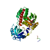 5vivC 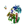 4xtqS C: citing same article ( S: Starting model for refinement |
|---|---|
| Similar structure data |


-Links
 PDBj
PDBj


- Assembly
| Deposited unit | 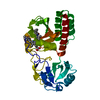
| ||||||||
|---|---|---|---|---|---|---|---|---|---|
| 1 |
| ||||||||
| Unit cell |
|
-Components
| #1: Protein | Mass: 34556.941 Da / Num. of mol.: 1 Source method: isolated from a genetically manipulated source Source: (gene. exp.) Rhodopseudomonas palustris (phototrophic)Production host: Escherichia coli (E. coli) / References: UniProt: B3Q7C0*PLUS |
|---|---|
| #2: Chemical | ChemComp-BLA /   Mass: 582.646 Da / Num. of mol.: 1 / Source method: obtained synthetically / Formula: C33H34N4O6 Mass: 582.646 Da / Num. of mol.: 1 / Source method: obtained synthetically / Formula: C33H34N4O6 |
| #3: Water | ChemComp-HOH / Water Mass: 18.015 Da / Num. of mol.: 280 / Source method: isolated from a natural source / Formula: H2O Mass: 18.015 Da / Num. of mol.: 280 / Source method: isolated from a natural source / Formula: H2O |
-Experimental details
-Experiment
| Experiment | Method: X-RAY DIFFRACTION / Number of used crystals: 1 |
|---|
- Sample preparation
| Crystal | Density Matthews: 1.79 Å3/Da / Density % sol: 31.4 % |
|---|---|
| Crystal grow | Temperature: 293 K / Method: vapor diffusion, hanging drop Details: 0.08M Na Citrate pH 5.0, 24% v/v Jeffamine ED-2001 pH 7.0 0.4% n-octyl-beta-D-glucopyranoside |
-Data collection
| Diffraction | Mean temperature: 100 K | ||||||||||||||||||||||||||||||||||||||||||||||||||||||||||||||||||||||||||||||||||||||||
|---|---|---|---|---|---|---|---|---|---|---|---|---|---|---|---|---|---|---|---|---|---|---|---|---|---|---|---|---|---|---|---|---|---|---|---|---|---|---|---|---|---|---|---|---|---|---|---|---|---|---|---|---|---|---|---|---|---|---|---|---|---|---|---|---|---|---|---|---|---|---|---|---|---|---|---|---|---|---|---|---|---|---|---|---|---|---|---|---|---|
| Diffraction source | Source: SYNCHROTRON / Site: APS  / Beamline: 22-BM / Wavelength: 1 Å / Beamline: 22-BM / Wavelength: 1 Å | ||||||||||||||||||||||||||||||||||||||||||||||||||||||||||||||||||||||||||||||||||||||||
| Detector | Type: MARMOSAIC 225 mm CCD / Detector: CCD / Date: Dec 1, 2016 | ||||||||||||||||||||||||||||||||||||||||||||||||||||||||||||||||||||||||||||||||||||||||
| Radiation | Protocol: SINGLE WAVELENGTH / Monochromatic (M) / Laue (L): M / Scattering type: x-ray | ||||||||||||||||||||||||||||||||||||||||||||||||||||||||||||||||||||||||||||||||||||||||
| Radiation wavelength | Wavelength: 1 Å / Relative weight: 1 | ||||||||||||||||||||||||||||||||||||||||||||||||||||||||||||||||||||||||||||||||||||||||
| Reflection | Resolution: 1.35→29.93 Å / Num. obs: 53575 / % possible obs: 99.9 % / Redundancy: 3.7 % / Rmerge(I) obs: 0.054 / Rpim(I) all: 0.033 / Rrim(I) all: 0.063 / Χ2: 0.898 / Net I/σ(I): 13.6 / Num. measured all: 197471 | ||||||||||||||||||||||||||||||||||||||||||||||||||||||||||||||||||||||||||||||||||||||||
| Reflection shell | Diffraction-ID: 1
|
- Processing
| Software |
| |||||||||||||||||||||||||||||||||||||||||||||||||||||||||||||||||||||||||||
|---|---|---|---|---|---|---|---|---|---|---|---|---|---|---|---|---|---|---|---|---|---|---|---|---|---|---|---|---|---|---|---|---|---|---|---|---|---|---|---|---|---|---|---|---|---|---|---|---|---|---|---|---|---|---|---|---|---|---|---|---|---|---|---|---|---|---|---|---|---|---|---|---|---|---|---|---|
| Refinement | Method to determine structure: MOLECULAR REPLACEMENT Starting model: 4XTQ Resolution: 1.35→29.93 Å / Cor.coef. Fo:Fc: 0.983 / Cor.coef. Fo:Fc free: 0.968 / SU B: 1.979 / SU ML: 0.036 / Cross valid method: THROUGHOUT / σ(F): 0 / ESU R: 0.053 / ESU R Free: 0.055 Details: HYDROGENS HAVE BEEN ADDED IN THE RIDING POSITIONS U VALUES : REFINED INDIVIDUALLY
| |||||||||||||||||||||||||||||||||||||||||||||||||||||||||||||||||||||||||||
| Solvent computation | Ion probe radii: 0.8 Å / Shrinkage radii: 0.8 Å / VDW probe radii: 1.2 Å | |||||||||||||||||||||||||||||||||||||||||||||||||||||||||||||||||||||||||||
| Displacement parameters | Biso max: 101.64 Å2 / Biso mean: 20.485 Å2 / Biso min: 8.87 Å2
| |||||||||||||||||||||||||||||||||||||||||||||||||||||||||||||||||||||||||||
| Refinement step | Cycle: final / Resolution: 1.35→29.93 Å
| |||||||||||||||||||||||||||||||||||||||||||||||||||||||||||||||||||||||||||
| Refine LS restraints |
| |||||||||||||||||||||||||||||||||||||||||||||||||||||||||||||||||||||||||||
| LS refinement shell | Resolution: 1.351→1.386 Å / Rfactor Rfree error: 0 / Total num. of bins used: 20
|
