 Movie
Movie Controller

[English] 日本語
 Yorodumi
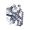
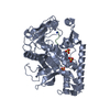
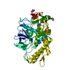
- PDB-5vgx: Structure of the C. botulinum neurotoxin serotype A with Hg bound -
+ Open data

- Basic information
| Entry | Database: PDB / ID: 5vgx | ||||||
|---|---|---|---|---|---|---|---|
| Title | Structure of the C. botulinum neurotoxin serotype A with Hg bound | ||||||
 Components Components | Botulinum neurotoxin type A | ||||||
 Keywords Keywords |  TOXIN / botulinum / neurotoxin / metalloprotease TOXIN / botulinum / neurotoxin / metalloprotease | ||||||
| Function / homology |  Function and homology information Function and homology informationhost cell junction / negative regulation of neurotransmitter secretion / bontoxilysin / host cell presynaptic membrane / host cell cytoplasmic vesicle / host cell cytosol / protein transmembrane transporter activity / metalloendopeptidase activity / toxin activity / membrane => GO:0016020 ...host cell junction / negative regulation of neurotransmitter secretion / bontoxilysin / host cell presynaptic membrane / host cell cytoplasmic vesicle / host cell cytosol / protein transmembrane transporter activity / metalloendopeptidase activity / toxin activity / membrane => GO:0016020 / host cell plasma membrane / proteolysis / zinc ion binding / extracellular region / membraneSimilarity search - Function | ||||||
| Biological species |   Clostridium botulinum (bacteria) Clostridium botulinum (bacteria) | ||||||
| Method | X-RAY DIFFRACTION / SYNCHROTRON / MOLECULAR REPLACEMENT / Resolution: 2.15 Å | ||||||
 Authors Authors | Carolan, J.P. / Allen, K.N. | ||||||
| Funding support |  United States, 1items United States, 1items
| ||||||
 Citation Citation | Journal: J. Am. Chem. Soc. / Year: 2017 Title: Metal Ions Effectively Ablate the Action of Botulinum Neurotoxin A. Authors: Bremer, P.T. / Pellett, S. / Carolan, J.P. / Tepp, W.H. / Eubanks, L.M. / Allen, K.N. / Johnson, E.A. / Janda, K.D. | ||||||
| History |
|
- Structure visualization
| Structure viewer | Molecule: MolmilJmol/JSmol |
|---|
- Downloads & links
-Download
| PDBx/mmCIF format | 5vgx.cif.gz | 189.5 KB | Display | PDBx/mmCIF format |
|---|---|---|---|---|
| PDB format | pdb5vgx.ent.gz | 148.4 KB | Display | PDB format |
| PDBx/mmJSON format | 5vgx.json.gz | Tree view | PDBx/mmJSON format | |
| Others |  Other downloads Other downloads |
-Validation report
| Arichive directory | https://data.pdbj.org/pub/pdb/validation_reports/vg/5vgxftp://data.pdbj.org/pub/pdb/validation_reports/vg/5vgx | HTTPS FTP |
|---|
-Related structure data
| Related structure data |  5vgvC  3bokS C: citing same article ( S: Starting model for refinement |
|---|---|
| Similar structure data |

-Links
 PDBj
PDBj


- Assembly
| Deposited unit | 
| ||||||||
|---|---|---|---|---|---|---|---|---|---|
| 1 |
| ||||||||
| Unit cell |
|
-Components
| #1: Protein | Mass: 50602.047 Da / Num. of mol.: 1 Source method: isolated from a genetically manipulated source Details: Polyhistidine-tagged botulinum neurotoxin A light chain Source: (gene. exp.) Clostridium botulinum (bacteria) / Gene: botA, atx, bna / Production host: Escherichia coli (E. coli) / Strain (production host): BL21(DE3)References: UniProt: P10845, UniProt: P0DPI1*PLUS, bontoxilysin |
|---|---|
| #2: Chemical | ChemComp-HG / Mercury (element)  Mass: 200.590 Da / Num. of mol.: 1 / Source method: obtained synthetically / Formula: Hg Mass: 200.590 Da / Num. of mol.: 1 / Source method: obtained synthetically / Formula: Hg |
| #3: Chemical | ChemComp-ACT / Acetate  Mass: 59.044 Da / Num. of mol.: 1 / Source method: obtained synthetically / Formula: C2H3O2 Mass: 59.044 Da / Num. of mol.: 1 / Source method: obtained synthetically / Formula: C2H3O2 |
| #4: Water | ChemComp-HOH / Water Mass: 18.015 Da / Num. of mol.: 98 / Source method: isolated from a natural source / Formula: H2O Mass: 18.015 Da / Num. of mol.: 98 / Source method: isolated from a natural source / Formula: H2O |
-Experimental details
-Experiment
| Experiment | Method: X-RAY DIFFRACTION / Number of used crystals: 1 |
|---|
- Sample preparation
| Crystal | Density Matthews: 2.1 Å3/Da / Density % sol: 41.31 % |
|---|---|
| Crystal grow | Temperature: 290 K / Method: vapor diffusion, hanging drop / pH: 6.5 Details: PEG8000 8%, Calcium Acetate 0.2 M, Sodium Cacodylate 0.1 M |
-Data collection
| Diffraction | Mean temperature: 100 K / Ambient temp details: N2 Stream |
|---|---|
| Diffraction source | Source: SYNCHROTRON / Site: SSRL / Beamline: BL9-2 / Wavelength: 0.99164 Å |
| Detector | Type: DECTRIS PILATUS 6M / Detector: PIXEL / Date: Feb 4, 2017 / Details: Mirror: Rh coated flat, toroidal focusing |
| Radiation | Monochromator: Si(111) and Si(220) double crystal / Protocol: SINGLE WAVELENGTH / Monochromatic (M) / Laue (L): M / Scattering type: x-ray |
| Radiation wavelength | Wavelength: 0.99164 Å / Relative weight: 1 |
| Reflection | Resolution: 2.15→27.5393 Å / Num. obs: 22337 / % possible obs: 98 % / Redundancy: 4.8 % / Biso Wilson estimate: 24.46 Å2 / CC1/2: 0.98 / Rmerge(I) obs: 0.1515 / Χ2: 1.084 / Net I/σ(I): 8.11 |
| Reflection shell | Resolution: 2.15→2.227 Å / Redundancy: 3.9 % / Rmerge(I) obs: 0.3144 / Mean I/σ(I) obs: 2.7 / Num. unique obs: 2145 / CC1/2: 0.909 / Χ2: 0.371 / % possible all: 95 |
- Processing
| Software |
| |||||||||||||||||||||||||||||||||||||||||||||||||||||||||||||||||||||||||||||||||||||||||||||||||||||||||||||||||||||||||||||||||||||
|---|---|---|---|---|---|---|---|---|---|---|---|---|---|---|---|---|---|---|---|---|---|---|---|---|---|---|---|---|---|---|---|---|---|---|---|---|---|---|---|---|---|---|---|---|---|---|---|---|---|---|---|---|---|---|---|---|---|---|---|---|---|---|---|---|---|---|---|---|---|---|---|---|---|---|---|---|---|---|---|---|---|---|---|---|---|---|---|---|---|---|---|---|---|---|---|---|---|---|---|---|---|---|---|---|---|---|---|---|---|---|---|---|---|---|---|---|---|---|---|---|---|---|---|---|---|---|---|---|---|---|---|---|---|---|
| Refinement | Method to determine structure: MOLECULAR REPLACEMENT Starting model: 3BOK Resolution: 2.15→27.537 Å / SU ML: 0.26 / Cross valid method: FREE R-VALUE / σ(F): 1.39 / Phase error: 26.81 Details: Refinement performed utilizing XYZ coordinate, real space, TLS, anomalous groups, and individual B-factor parameters.
| |||||||||||||||||||||||||||||||||||||||||||||||||||||||||||||||||||||||||||||||||||||||||||||||||||||||||||||||||||||||||||||||||||||
| Solvent computation | Shrinkage radii: 0.9 Å / VDW probe radii: 1.11 Å | |||||||||||||||||||||||||||||||||||||||||||||||||||||||||||||||||||||||||||||||||||||||||||||||||||||||||||||||||||||||||||||||||||||
| Refinement step | Cycle: LAST / Resolution: 2.15→27.537 Å
| |||||||||||||||||||||||||||||||||||||||||||||||||||||||||||||||||||||||||||||||||||||||||||||||||||||||||||||||||||||||||||||||||||||
| Refine LS restraints |
| |||||||||||||||||||||||||||||||||||||||||||||||||||||||||||||||||||||||||||||||||||||||||||||||||||||||||||||||||||||||||||||||||||||
| LS refinement shell |
| |||||||||||||||||||||||||||||||||||||||||||||||||||||||||||||||||||||||||||||||||||||||||||||||||||||||||||||||||||||||||||||||||||||
| Refinement TLS params. | Method: refined / Origin x: 5.1333 Å / Origin y: -0.583 Å / Origin z: 80.5388 Å
| |||||||||||||||||||||||||||||||||||||||||||||||||||||||||||||||||||||||||||||||||||||||||||||||||||||||||||||||||||||||||||||||||||||
| Refinement TLS group | Selection details: all |
