- PDB-5vaz: Crystal structure of a DNA primase domain from Pseudomonas aeruginosa -
+
Open data
ID or keywords:
Loading...
-
Basic information
Entry
Database: PDB / ID: 5vaz
Title
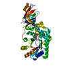
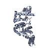
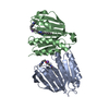
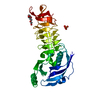
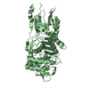
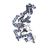
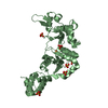
Crystal structure of a DNA primase domain from Pseudomonas aeruginosa
Components
DNA primasePrimase
Keywords
TRANSFERASE / structural genomics / DNA-binding / primase / truncation / Seattle Structural Genomics Center for Infectious Disease / SSGCID
Function / homology
Function and homology information
primosome complex / DNA primase activity / DNA replication, synthesis of primer / Transferases; Transferring phosphorus-containing groups; Nucleotidyltransferases / DNA binding / zinc ion binding / cytoplasm Similarity search - Function
DnaG, RNA polymerase domain, helical bundle / DNA primase, catalytic core, N-terminal domain / DNA primase DnaG, DnaB-binding domain / DNA primase DnaG DnaB-binding / DNA primase DnaG DnaB-binding / DNA primase DNAg catalytic core, N-terminal domain / Pheromone ER-1 / DNA primase, DnaB-helicase binding domain / DnaB-helicase binding domain of primase / Toprim domain ...DnaG, RNA polymerase domain, helical bundle / DNA primase, catalytic core, N-terminal domain / DNA primase DnaG, DnaB-binding domain / DNA primase DnaG DnaB-binding / DNA primase DnaG DnaB-binding / DNA primase DNAg catalytic core, N-terminal domain / Pheromone ER-1 / DNA primase, DnaB-helicase binding domain / DnaB-helicase binding domain of primase / Toprim domain / Dna Topoisomerase Vi A Subunit; Chain: A, domain 2 / Dna Topoisomerase Vi A Subunit; Chain: A, domain 2 - #10 / Zinc finger, CHC2-type / DNA primase, DnaG / DNA primase, catalytic core, N-terminal / DNA primase DnaG, bacteria / Bacterial DnaG primase, TOPRIM domain / DNA Primase, CHC2-type zinc finger / DNA primase, catalytic core, N-terminal domain superfamily / CHC2 zinc finger / DNA primase catalytic core, N-terminal domain / zinc finger / DNA helicase DnaB, N-terminal/DNA primase DnaG, C-terminal / TOPRIM / Toprim domain profile. / TOPRIM domain / Alpha-Beta Complex / Up-down Bundle / 3-Layer(aba) Sandwich / Mainly Alpha / Alpha Beta Similarity search - Domain/homology
Mass: 18.015 Da / Num. of mol.: 202 / Source method: isolated from a natural source / Formula: H2O
-
Experimental details
-
Experiment
Experiment
Method: X-RAY DIFFRACTION / Number of used crystals: 1
-
Sample preparation
Crystal
Density Matthews: 3.6 Å3/Da / Density % sol: 65.87 %
Crystal grow
Temperature: 289 K / Method: vapor diffusion, sitting drop / pH: 8.5 Details: PsaeA.00153.a.B7.PS38108 at 18.8 mg/mL against Top96 screen condition A4: 0.1 M Tris pH 8.5, 2 M ammonium sulfate, cryo-protected with 20% EG, crystal tracking ID 28610a4
In the structure databanks used in Yorodumi, some data are registered as the other names, "COVID-19 virus" and "2019-nCoV". Here are the details of the virus and the list of structure data.
Jan 31, 2019. EMDB accession codes are about to change! (news from PDBe EMDB page)
EMDB accession codes are about to change! (news from PDBe EMDB page)
The allocation of 4 digits for EMDB accession codes will soon come to an end. Whilst these codes will remain in use, new EMDB accession codes will include an additional digit and will expand incrementally as the available range of codes is exhausted. The current 4-digit format prefixed with “EMD-” (i.e. EMD-XXXX) will advance to a 5-digit format (i.e. EMD-XXXXX), and so on. It is currently estimated that the 4-digit codes will be depleted around Spring 2019, at which point the 5-digit format will come into force.
The EM Navigator/Yorodumi systems omit the EMD- prefix.
Related info.:Q: What is EMD? / ID/Accession-code notation in Yorodumi/EM Navigator
Yorodumi is a browser for structure data from EMDB, PDB, SASBDB, etc.
This page is also the successor to EM Navigator detail page, and also detail information page/front-end page for Omokage search.
The word "yorodu" (or yorozu) is an old Japanese word meaning "ten thousand". "mi" (miru) is to see.
Related info.:EMDB / PDB / SASBDB / Comparison of 3 databanks / Yorodumi Search / Aug 31, 2016. New EM Navigator & Yorodumi / Yorodumi Papers / Jmol/JSmol / Function and homology information / Changes in new EM Navigator and Yorodumi
 Movie
Movie Controller

 Yorodumi
 Open data
 Basic information
 Components
 Primase
  Keywords
 Function and homology information

 Authors
 Citation
 Structure visualization
 Downloads & links
 Other downloads


 PDBj
PDBj

 Assembly
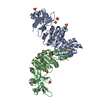

 Mass: 96.063 Da / Num. of mol.: 15 / Source method: obtained synthetically / Formula: SO4
 Mass: 18.015 Da / Num. of mol.: 202 / Source method: isolated from a natural source / Formula: H2O
 Sample preparation
 / Beamline: 21-ID-F / Wavelength: 0.97872 Å
 Processing