 Movie
Movie Controller

[English] 日本語
 Yorodumi
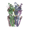
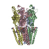
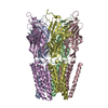
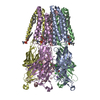
- PDB-5v6o: Crystal Structure of the highly open channel-stabilized mutant G-... -
+ Open data

- Basic information
| Entry | Database: PDB / ID: 5v6o | ||||||
|---|---|---|---|---|---|---|---|
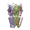
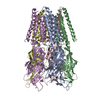
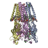
| Title | Crystal Structure of the highly open channel-stabilized mutant G-2'I + I9'A of GLIC | ||||||
 Components Components | Proton-gated ion channel | ||||||
 Keywords Keywords |  membrane protein / metal transport / GLIC / Cys-loop receptors / ion channel / GluCl / ELIC membrane protein / metal transport / GLIC / Cys-loop receptors / ion channel / GluCl / ELIC | ||||||
| Function / homology |  Function and homology informationsodium channel activity / extracellular ligand-gated monoatomic ion channel activity / transmembrane transporter complex / potassium channel activity / regulation of membrane potential / transmembrane signaling receptor activity / neuron projection / signal transduction / identical protein binding / plasma membrane Function and homology informationsodium channel activity / extracellular ligand-gated monoatomic ion channel activity / transmembrane transporter complex / potassium channel activity / regulation of membrane potential / transmembrane signaling receptor activity / neuron projection / signal transduction / identical protein binding / plasma membraneSimilarity search - Function | ||||||
| Biological species |  Gloeobacter violaceus (bacteria) Gloeobacter violaceus (bacteria) | ||||||
| Method | X-RAY DIFFRACTION / SYNCHROTRON / MOLECULAR REPLACEMENT / Resolution: 3.121 Å | ||||||
 Authors Authors | Gonzalez-Gutierrez, G. / Grosman, C. | ||||||
| Funding support |  United States, 1items United States, 1items
| ||||||
 Citation Citation | Journal: J. Gen. Physiol. / Year: 2017 Title: Chasing the open-state structure of pentameric ligand-gated ion channels. Authors: Gonzalez-Gutierrez, G. / Wang, Y. / Cymes, G.D. / Tajkhorshid, E. / Grosman, C. | ||||||
| History |
|
- Structure visualization
| Structure viewer | Molecule: MolmilJmol/JSmol |
|---|
- Downloads & links
-Download
| PDBx/mmCIF format | 5v6o.cif.gz | 318.4 KB | Display | PDBx/mmCIF format |
|---|---|---|---|---|
| PDB format | pdb5v6o.ent.gz | 259.3 KB | Display | PDB format |
| PDBx/mmJSON format | 5v6o.json.gz | Tree view | PDBx/mmJSON format | |
| Others |  Other downloads Other downloads |
-Validation report
| Arichive directory | https://data.pdbj.org/pub/pdb/validation_reports/v6/5v6oftp://data.pdbj.org/pub/pdb/validation_reports/v6/5v6o | HTTPS FTP |
|---|
-Related structure data
| Related structure data | 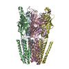 5v6nC  3ehzS C: citing same article ( S: Starting model for refinement |
|---|---|
| Similar structure data |


-Links
 PDBj
PDBj

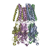
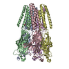
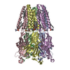
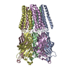
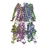
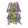
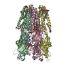
- Assembly
| Deposited unit | 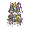
| ||||||||||||||||||||||||||||||||||||||||||||||||||||||
|---|---|---|---|---|---|---|---|---|---|---|---|---|---|---|---|---|---|---|---|---|---|---|---|---|---|---|---|---|---|---|---|---|---|---|---|---|---|---|---|---|---|---|---|---|---|---|---|---|---|---|---|---|---|---|---|
| 1 |
| ||||||||||||||||||||||||||||||||||||||||||||||||||||||
| Unit cell |
| ||||||||||||||||||||||||||||||||||||||||||||||||||||||
| Noncrystallographic symmetry (NCS) | NCS domain:
NCS domain segments:
|
-Components
| #1: Protein | Mass: 35504.910 Da / Num. of mol.: 5 / Fragment: unp residues 50-359 / Mutation: C27S, K33C Source method: isolated from a genetically manipulated source Source: (gene. exp.) Gloeobacter violaceus (strain PCC 7421) (bacteria)Strain: PCC 7421 / Gene: glvI, glr4197 / Production host: Escherichia coli (E. coli) / References: UniProt: Q7NDN8#2: Sugar | ChemComp-LMT /   Type: D-saccharide / Mass: 510.615 Da / Num. of mol.: 6 / Source method: obtained synthetically / Formula: C24H46O11 / Comment: detergent*YM Type: D-saccharide / Mass: 510.615 Da / Num. of mol.: 6 / Source method: obtained synthetically / Formula: C24H46O11 / Comment: detergent*YM#3: Chemical | ChemComp-CL / Chloride  Mass: 35.453 Da / Num. of mol.: 5 / Source method: obtained synthetically / Formula: Cl Mass: 35.453 Da / Num. of mol.: 5 / Source method: obtained synthetically / Formula: Cl#4: Chemical | ChemComp-NA / |   Mass: 22.990 Da / Num. of mol.: 1 / Source method: obtained synthetically / Formula: Na Mass: 22.990 Da / Num. of mol.: 1 / Source method: obtained synthetically / Formula: Na#5: Chemical | ChemComp-SO4 / Sulfate  Mass: 96.063 Da / Num. of mol.: 10 / Source method: obtained synthetically / Formula: SO4 Mass: 96.063 Da / Num. of mol.: 10 / Source method: obtained synthetically / Formula: SO4 |
|---|
-Experimental details
-Experiment
| Experiment | Method: X-RAY DIFFRACTION / Number of used crystals: 1 |
|---|
- Sample preparation
| Crystal | Density Matthews: 5.26 Å3/Da / Density % sol: 76.6 % |
|---|---|
| Crystal grow | Temperature: 277 K / Method: vapor diffusion, hanging drop Details: Sodium Acetate 50 mM pH 3.9-4.2 Ammonium Sulfate 200-250 mM PEG 4000 10-12% PH range: 3.9-4.2 |
-Data collection
| Diffraction | Mean temperature: 100 K | |||||||||||||||||||||
|---|---|---|---|---|---|---|---|---|---|---|---|---|---|---|---|---|---|---|---|---|---|---|
| Diffraction source | Source: SYNCHROTRON / Site: APS / Beamline: 21-ID-G / Wavelength: 0.97857 Å | |||||||||||||||||||||
| Detector | Type: MAR scanner 300 mm plate / Detector: IMAGE PLATE / Date: Aug 4, 2013 | |||||||||||||||||||||
| Radiation | Protocol: SINGLE WAVELENGTH / Monochromatic (M) / Laue (L): M / Scattering type: x-ray | |||||||||||||||||||||
| Radiation wavelength | Wavelength: 0.97857 Å / Relative weight: 1 | |||||||||||||||||||||
| Reflection | Resolution: 3.121→105.979 Å / Num. obs: 65357 / % possible obs: 99.9 % / Redundancy: 4.2 % / Biso Wilson estimate: 66.78 Å2 / CC1/2: 0.995 / Rmerge(I) obs: 0.162 / Rpim(I) all: 0.089 / Rrim(I) all: 0.186 / Net I/σ(I): 8.7 / Num. measured all: 276516 | |||||||||||||||||||||
| Reflection shell | Diffraction-ID: 1
|
- Processing
| Software |
| ||||||||||||||||||||||||||||||||||||||||||||||||||||||||||||||||||||||||||||||||||||||||||||||||||||||||||||||||||||||||||||||||||||||||||||||||||||||||||||||||||||||||
|---|---|---|---|---|---|---|---|---|---|---|---|---|---|---|---|---|---|---|---|---|---|---|---|---|---|---|---|---|---|---|---|---|---|---|---|---|---|---|---|---|---|---|---|---|---|---|---|---|---|---|---|---|---|---|---|---|---|---|---|---|---|---|---|---|---|---|---|---|---|---|---|---|---|---|---|---|---|---|---|---|---|---|---|---|---|---|---|---|---|---|---|---|---|---|---|---|---|---|---|---|---|---|---|---|---|---|---|---|---|---|---|---|---|---|---|---|---|---|---|---|---|---|---|---|---|---|---|---|---|---|---|---|---|---|---|---|---|---|---|---|---|---|---|---|---|---|---|---|---|---|---|---|---|---|---|---|---|---|---|---|---|---|---|---|---|---|---|---|---|
| Refinement | Method to determine structure: MOLECULAR REPLACEMENT Starting model: 3EHZ Resolution: 3.121→70.443 Å / SU ML: 0.51 / Cross valid method: FREE R-VALUE / σ(F): 1.34 / Phase error: 33.59
| ||||||||||||||||||||||||||||||||||||||||||||||||||||||||||||||||||||||||||||||||||||||||||||||||||||||||||||||||||||||||||||||||||||||||||||||||||||||||||||||||||||||||
| Solvent computation | Shrinkage radii: 0.9 Å / VDW probe radii: 1.11 Å | ||||||||||||||||||||||||||||||||||||||||||||||||||||||||||||||||||||||||||||||||||||||||||||||||||||||||||||||||||||||||||||||||||||||||||||||||||||||||||||||||||||||||
| Displacement parameters | Biso max: 142.87 Å2 / Biso mean: 67.4838 Å2 / Biso min: 38.1 Å2 | ||||||||||||||||||||||||||||||||||||||||||||||||||||||||||||||||||||||||||||||||||||||||||||||||||||||||||||||||||||||||||||||||||||||||||||||||||||||||||||||||||||||||
| Refinement step | Cycle: final / Resolution: 3.121→70.443 Å
| ||||||||||||||||||||||||||||||||||||||||||||||||||||||||||||||||||||||||||||||||||||||||||||||||||||||||||||||||||||||||||||||||||||||||||||||||||||||||||||||||||||||||
| Refine LS restraints |
| ||||||||||||||||||||||||||||||||||||||||||||||||||||||||||||||||||||||||||||||||||||||||||||||||||||||||||||||||||||||||||||||||||||||||||||||||||||||||||||||||||||||||
| Refine LS restraints NCS |
| ||||||||||||||||||||||||||||||||||||||||||||||||||||||||||||||||||||||||||||||||||||||||||||||||||||||||||||||||||||||||||||||||||||||||||||||||||||||||||||||||||||||||
| LS refinement shell | Refine-ID: X-RAY DIFFRACTION / Rfactor Rfree error: 0 / Total num. of bins used: 23
|
