 Movie
Movie Controller

[English] 日本語
 Yorodumi
- PDB-5v6f: Crystal Structure of the Second beta-Prism Domain of RbmC from V.... -
+ Open data

- Basic information
| Entry | Database: PDB / ID: 5v6f | |||||||||
|---|---|---|---|---|---|---|---|---|---|---|
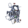
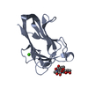
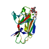
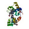
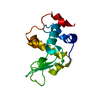
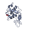
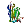
| Title | Crystal Structure of the Second beta-Prism Domain of RbmC from V. cholerae bound to Mannotriose | |||||||||
 Components Components | Hemolysin-related protein | |||||||||
 Keywords Keywords | SUGAR BINDING PROTEIN /  lectin / beta-prism / glycan-binding domain / mannotriose lectin / beta-prism / glycan-binding domain / mannotriose | |||||||||
| Function / homology |  Function and homology information Function and homology informationMetalloprotease StcE, C-terminal / Beta/Gamma crystallin / Hemolysin, beta-prism lectin / Beta-prism lectin / FG-GAP-like repeat / Jacalin-like lectin domain / Aligned Prism / Vitelline Membrane Outer Layer Protein I, subunit A / Jacalin-like lectin domain superfamily / FG-GAP repeat ...Metalloprotease StcE, C-terminal / Beta/Gamma crystallin / Hemolysin, beta-prism lectin / Beta-prism lectin / FG-GAP-like repeat / Jacalin-like lectin domain / Aligned Prism / Vitelline Membrane Outer Layer Protein I, subunit A / Jacalin-like lectin domain superfamily / FG-GAP repeat / Integrin alpha, N-terminal / EF-Hand 1, calcium-binding site / EF-hand calcium-binding domain. / Mainly BetaSimilarity search - Domain/homology | |||||||||
| Biological species |  Vibrio cholerae serotype O1 (bacteria) Vibrio cholerae serotype O1 (bacteria) | |||||||||
| Method | X-RAY DIFFRACTION / MOLECULAR REPLACEMENT / molecular replacement / Resolution: 1.5 Å | |||||||||
| Model details | apo form | |||||||||
 Authors Authors | De, S. / Kaus, K. / Sinclair, S. / Case, B.C. / Olson, R. | |||||||||
| Funding support |  United States, 1items United States, 1items
| |||||||||
 Citation Citation | Journal: PLoS Pathog. / Year: 2018 Title: Structural basis of mammalian glycan targeting by Vibrio cholerae cytolysin and biofilm proteins. Authors: De, S. / Kaus, K. / Sinclair, S. / Case, B.C. / Olson, R. | |||||||||
| History |
|
- Structure visualization
| Structure viewer | Molecule: MolmilJmol/JSmol |
|---|
- Downloads & links
-Download
| PDBx/mmCIF format | 5v6f.cif.gz | 71.9 KB | Display | PDBx/mmCIF format |
|---|---|---|---|---|
| PDB format | pdb5v6f.ent.gz | 50.7 KB | Display | PDB format |
| PDBx/mmJSON format | 5v6f.json.gz | Tree view | PDBx/mmJSON format | |
| Others |  Other downloads Other downloads |
-Validation report
| Arichive directory | https://data.pdbj.org/pub/pdb/validation_reports/v6/5v6fftp://data.pdbj.org/pub/pdb/validation_reports/v6/5v6f | HTTPS FTP |
|---|
-Related structure data
| Related structure data | 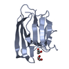 5v6cC 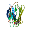 5v6kC  1xezS C: citing same article ( S: Starting model for refinement |
|---|---|
| Similar structure data |


-Links
 PDBj
PDBj
- Assembly
| Deposited unit | 
| ||||||||
|---|---|---|---|---|---|---|---|---|---|
| 1 |
| ||||||||
| Unit cell |
|
-Components
| #1: Protein | Mass: 14932.883 Da / Num. of mol.: 1 / Fragment: lectin domain, UNP residues 823-957 Source method: isolated from a genetically manipulated source Source: (gene. exp.) Vibrio cholerae serotype O1 (strain ATCC 39315 / El Tor Inaba N16961) (bacteria)Strain: ATCC 39315 / El Tor Inaba N16961 / Gene: VC_0930 / Plasmid: pNGFP-BC / Production host: Escherichia coli (E. coli) / Strain (production host): NEB T7 Express / References: UniProt: Q9KTH2 | ||
|---|---|---|---|
| #2: Polysaccharide | alpha-D-mannopyranose-(1-3)-[alpha-D-mannopyranose-(1-6)]beta-D-mannopyranose / Mass: 504.438 Da / Num. of mol.: 1 Source method: isolated from a genetically manipulated source | ||
| #3: Chemical | Sulfate  Mass: 96.063 Da / Num. of mol.: 2 / Source method: obtained synthetically / Formula: SO4 Mass: 96.063 Da / Num. of mol.: 2 / Source method: obtained synthetically / Formula: SO4#4: Water | ChemComp-HOH / | Water Mass: 18.015 Da / Num. of mol.: 237 / Source method: isolated from a natural source / Formula: H2O Mass: 18.015 Da / Num. of mol.: 237 / Source method: isolated from a natural source / Formula: H2O |
-Experimental details
-Experiment
| Experiment | Method: X-RAY DIFFRACTION / Number of used crystals: 1 |
|---|
- Sample preparation
| Crystal | Density Matthews: 2 Å3/Da / Density % sol: 38.44 % / Mosaicity: 0.83 ° |
|---|---|
| Crystal grow | Temperature: 295 K / Method: vapor diffusion, hanging drop / pH: 4.6 Details: 0.1 M sodium acetate, 2 M sodium sulfate, 2 mM mannotriose |
-Data collection
| Diffraction | Mean temperature: 100 K | |||||||||||||||||||||||||||||||||||||||||||||||||||||||||||||||||||||||||||||||||||||||||||||||||||||||||||||||||||||||||
|---|---|---|---|---|---|---|---|---|---|---|---|---|---|---|---|---|---|---|---|---|---|---|---|---|---|---|---|---|---|---|---|---|---|---|---|---|---|---|---|---|---|---|---|---|---|---|---|---|---|---|---|---|---|---|---|---|---|---|---|---|---|---|---|---|---|---|---|---|---|---|---|---|---|---|---|---|---|---|---|---|---|---|---|---|---|---|---|---|---|---|---|---|---|---|---|---|---|---|---|---|---|---|---|---|---|---|---|---|---|---|---|---|---|---|---|---|---|---|---|---|---|---|
| Diffraction source | Source: SEALED TUBE / Type: OXFORD DIFFRACTION NOVA / Wavelength: 1.54 Å | |||||||||||||||||||||||||||||||||||||||||||||||||||||||||||||||||||||||||||||||||||||||||||||||||||||||||||||||||||||||||
| Detector | Type: OXFORD ONYX CCD / Detector: CCD / Date: May 26, 2016 | |||||||||||||||||||||||||||||||||||||||||||||||||||||||||||||||||||||||||||||||||||||||||||||||||||||||||||||||||||||||||
| Radiation | Protocol: SINGLE WAVELENGTH / Monochromatic (M) / Laue (L): M / Scattering type: x-ray | |||||||||||||||||||||||||||||||||||||||||||||||||||||||||||||||||||||||||||||||||||||||||||||||||||||||||||||||||||||||||
| Radiation wavelength | Wavelength: 1.54 Å / Relative weight: 1 | |||||||||||||||||||||||||||||||||||||||||||||||||||||||||||||||||||||||||||||||||||||||||||||||||||||||||||||||||||||||||
| Reflection | Resolution: 1.5→16.112 Å / Num. obs: 18634 / % possible obs: 95.1 % / Redundancy: 2.6 % / Rpim(I) all: 0.056 / Rrim(I) all: 0.097 / Rsym value: 0.078 / Net I/av σ(I): 7.5 / Net I/σ(I): 7.7 / Num. measured all: 48096 | |||||||||||||||||||||||||||||||||||||||||||||||||||||||||||||||||||||||||||||||||||||||||||||||||||||||||||||||||||||||||
| Reflection shell | Diffraction-ID: 1
|
-Phasing
| Phasing | Method: molecular replacement | |||||||||
|---|---|---|---|---|---|---|---|---|---|---|
| Phasing MR | Model details: Phaser MODE: MR_AUTO
|
- Processing
| Software |
| ||||||||||||||||||||||||||||||||||||||||||||||||||||||||||||||||||||||||||||||||||||||||||||||||||
|---|---|---|---|---|---|---|---|---|---|---|---|---|---|---|---|---|---|---|---|---|---|---|---|---|---|---|---|---|---|---|---|---|---|---|---|---|---|---|---|---|---|---|---|---|---|---|---|---|---|---|---|---|---|---|---|---|---|---|---|---|---|---|---|---|---|---|---|---|---|---|---|---|---|---|---|---|---|---|---|---|---|---|---|---|---|---|---|---|---|---|---|---|---|---|---|---|---|---|---|
| Refinement | Method to determine structure: MOLECULAR REPLACEMENT Starting model: 1XEZ Resolution: 1.5→16.112 Å / SU ML: 0.14 / Cross valid method: THROUGHOUT / σ(F): 1 / Phase error: 19.19 / Stereochemistry target values: ML
| ||||||||||||||||||||||||||||||||||||||||||||||||||||||||||||||||||||||||||||||||||||||||||||||||||
| Solvent computation | Shrinkage radii: 0.9 Å / VDW probe radii: 1.11 Å / Solvent model: FLAT BULK SOLVENT MODEL | ||||||||||||||||||||||||||||||||||||||||||||||||||||||||||||||||||||||||||||||||||||||||||||||||||
| Displacement parameters | Biso max: 58.16 Å2 / Biso mean: 9.2837 Å2 / Biso min: 1.09 Å2 | ||||||||||||||||||||||||||||||||||||||||||||||||||||||||||||||||||||||||||||||||||||||||||||||||||
| Refinement step | Cycle: final / Resolution: 1.5→16.112 Å
| ||||||||||||||||||||||||||||||||||||||||||||||||||||||||||||||||||||||||||||||||||||||||||||||||||
| Refine LS restraints |
| ||||||||||||||||||||||||||||||||||||||||||||||||||||||||||||||||||||||||||||||||||||||||||||||||||
| LS refinement shell | Refine-ID: X-RAY DIFFRACTION / Rfactor Rfree error: 0 / Total num. of bins used: 13
|
