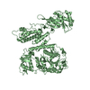
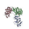
Entry Database : PDB / ID : 5v68Title Crystal structure of cell division protein FtsZ from Mycobacterium tuberculosis bounded via the T9 loop Cell division protein FtsZ Keywords / / / / / Function / homology Function Domain/homology Component
/ / / / / / / / / / / / / / / / / / / / / / / / / / / / / / / / / / / Biological species Mycobacterium tuberculosis (bacteria)Method / / / Resolution : 3.46 Å Authors Lazo, E.O. / Ojima, I. / Chowdhury, S.R. / Awasthi, D. / Jakoncic, J. Funding support Organization Grant number Country National Institutes of Health/National Institute Of Allergy and Infectious Diseases (NIH/NIAID) AIO78251 National Institutes of Health/National Institute Of Allergy and Infectious Diseases (NIH/NIAID) AIO82164 National Institutes of Health/National Institute of General Medical Sciences (NIH/NIGMS) GM-0080 Department of Energy (DOE, United States) DE-AC02-98CH10886 Office of Basic Energy Sciences DE-AC02-98CH10886 National Institutes of Health/National Center for Research Resources (NIH/NCRR) P41RR012408 National Institutes of Health/National Institute of General Medical Sciences (NIH/NIGMS) P41GM103473
Journal : Acta Crystallogr.,Sect.F / Year : 2019Title : Novel T9 loop conformation of filamenting temperature-sensitive mutant Z from Mycobacterium tuberculosis.Authors : Lazo, E.O. / Jakoncic, J. / RoyChowdhury, S. / Awasthi, D. / Ojima, I. History Deposition Mar 16, 2017 Deposition site / Processing site Revision 1.0 Mar 29, 2017 Provider / Type Revision 1.1 Sep 20, 2017 Group / Category / Item Revision 1.2 May 15, 2019 Group / Database references / Category / citation_authorItem _citation.country / _citation.journal_abbrev ... _citation.country / _citation.journal_abbrev / _citation.journal_id_ASTM / _citation.journal_id_CSD / _citation.journal_id_ISSN / _citation.journal_volume / _citation.page_first / _citation.page_last / _citation.pdbx_database_id_DOI / _citation.pdbx_database_id_PubMed / _citation.title / _citation.year / _citation_author.name Revision 1.3 Jul 17, 2019 Group / Refinement description / Category Item / _software.name / _software.versionRevision 1.4 Dec 4, 2019 Group / Category / Item Revision 1.5 Oct 4, 2023 Group / Database references / Refinement descriptionCategory chem_comp_atom / chem_comp_bond ... chem_comp_atom / chem_comp_bond / database_2 / pdbx_initial_refinement_model Item / _database_2.pdbx_database_accession
Show all Show less
 Movie
Movie Controller

 Yorodumi
 Open data
 Basic information
 Components
 Keywords
 Function and homology information

 Authors
 United States, 7items
  Citation
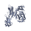
 Structure visualization
 Downloads & links
 Other downloads


 PDBj
PDBj


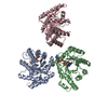
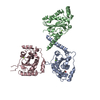
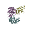
 Assembly

 Type: RNA linking / Mass: 443.201 Da / Num. of mol.: 2 / Source method: obtained synthetically / Formula: C10H15N5O11P2 / Comment: GDP, energy-carrying molecule*YM
 Mass: 94.971 Da / Num. of mol.: 2 / Source method: obtained synthetically / Formula: PO4
 Sample preparation
 Processing