 Movie
Movie Controller


+ Open data

- Basic information
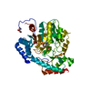
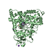
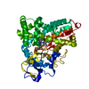
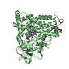
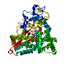
| Entry | Database: PDB / ID: 5uy6 | ||||||
|---|---|---|---|---|---|---|---|
| Title | Crystal Structure of the Human CAMKK2B | ||||||
 Components Components | Calcium/calmodulin-dependent protein kinase kinase 2 | ||||||
 Keywords Keywords | Transferase/Transferase Inhibitor /  transferase / protein kinase domain / Structural Genomics / Structural Genomics Consortium / SGC / Transferase-Transferase Inhibitor Complex transferase / protein kinase domain / Structural Genomics / Structural Genomics Consortium / SGC / Transferase-Transferase Inhibitor Complex | ||||||
| Function / homology |  Function and homology information Function and homology informationpositive regulation of autophagy of mitochondrion / Ca2+/calmodulin-dependent protein kinase / CAMKK-AMPK signaling cascade / regulation of protein kinase activity / calmodulin-dependent protein kinase activity / CREB1 phosphorylation through the activation of CaMKII/CaMKK/CaMKIV cascasde / CaMK IV-mediated phosphorylation of CREB / Activation of RAC1 downstream of NMDARs / Activation of AMPK downstream of NMDARs / calcium-mediated signaling ...positive regulation of autophagy of mitochondrion / Ca2+/calmodulin-dependent protein kinase / CAMKK-AMPK signaling cascade / regulation of protein kinase activity / calmodulin-dependent protein kinase activity / CREB1 phosphorylation through the activation of CaMKII/CaMKK/CaMKIV cascasde / CaMK IV-mediated phosphorylation of CREB / Activation of RAC1 downstream of NMDARs / Activation of AMPK downstream of NMDARs / calcium-mediated signaling / cellular response to reactive oxygen species / MAPK cascade / protein tyrosine kinase activity / protein autophosphorylation / calmodulin binding / neuron projection / positive regulation of protein phosphorylation / protein phosphorylation / protein serine kinase activity / protein serine/threonine kinase activity / calcium ion binding / positive regulation of DNA-templated transcription / nucleoplasm / ATP binding / cytosolSimilarity search - Function | ||||||
| Biological species |  Homo sapiens (human) Homo sapiens (human) | ||||||
| Method | X-RAY DIFFRACTION / SYNCHROTRON / MOLECULAR REPLACEMENT / Resolution: 1.7 Å | ||||||
 Authors Authors | Counago, R.M. / Dewry, D. / Bountra, C. / Arruda, P. / Edwards, A.M. / Gileadi, O. / Structural Genomics Consortium (SGC) | ||||||
| Funding support |  Brazil, 1items Brazil, 1items
| ||||||
 Citation Citation | Journal: To Be Published Title: Crystal Structure of the Human CAMKK2B Authors: Counago, R.M. / Dewry, D. / Bountra, C. / Arruda, P. / Edwards, A.M. / Gileadi, O. / Structural Genomics Consortium (SGC) | ||||||
| History |
|
- Structure visualization
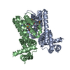
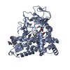
| Structure viewer | Molecule: MolmilJmol/JSmol |
|---|
- Downloads & links
-Download
| PDBx/mmCIF format | 5uy6.cif.gz | 238.3 KB | Display | PDBx/mmCIF format |
|---|---|---|---|---|
| PDB format | pdb5uy6.ent.gz | 191.6 KB | Display | PDB format |
| PDBx/mmJSON format | 5uy6.json.gz | Tree view | PDBx/mmJSON format | |
| Others |  Other downloads Other downloads |
-Validation report
| Arichive directory | https://data.pdbj.org/pub/pdb/validation_reports/uy/5uy6ftp://data.pdbj.org/pub/pdb/validation_reports/uy/5uy6 | HTTPS FTP |
|---|
-Related structure data
| Related structure data |  5uyjC  2zv2S C: citing same article ( S: Starting model for refinement |
|---|---|
| Similar structure data |


-Links
 PDBj
PDBj


- Assembly
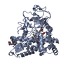
| Deposited unit | 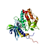
| ||||||||
|---|---|---|---|---|---|---|---|---|---|
| 1 |
| ||||||||
| Unit cell |
|
-Components
| #1: Protein | Mass: 33127.250 Da / Num. of mol.: 1 / Fragment: UNP residues 161-449 Source method: isolated from a genetically manipulated source Source: (gene. exp.) Homo sapiens (human) / Gene: CAMKK2, CAMKKB, KIAA0787 / Production host:  Escherichia coli (E. coli) / Strain (production host): BL21(DE3) / Variant (production host): R3 Escherichia coli (E. coli) / Strain (production host): BL21(DE3) / Variant (production host): R3References: UniProt: Q96RR4, Ca2+/calmodulin-dependent protein kinase |
|---|---|
| #2: Chemical | ChemComp-8R4 /   Mass: 383.439 Da / Num. of mol.: 1 / Source method: obtained synthetically / Formula: C25H21NO3 Mass: 383.439 Da / Num. of mol.: 1 / Source method: obtained synthetically / Formula: C25H21NO3 |
| #3: Water | ChemComp-HOH / Water Mass: 18.015 Da / Num. of mol.: 370 / Source method: isolated from a natural source / Formula: H2O Mass: 18.015 Da / Num. of mol.: 370 / Source method: isolated from a natural source / Formula: H2O |
-Experimental details
-Experiment
| Experiment | Method: X-RAY DIFFRACTION / Number of used crystals: 1 |
|---|
- Sample preparation
| Crystal | Density Matthews: 2.47 Å3/Da / Density % sol: 50.29 % |
|---|---|
| Crystal grow | Temperature: 293 K / Method: vapor diffusion, sitting drop / pH: 6.5 / Details: 25% PEG 3350; 0.1M Bis-Tris; 0.2M ammonium acetate |
-Data collection
| Diffraction | Mean temperature: 100 K |
|---|---|
| Diffraction source | Source: SYNCHROTRON / Site: APS  / Beamline: 24-ID-C / Wavelength: 0.9792 Å / Beamline: 24-ID-C / Wavelength: 0.9792 Å |
| Detector | Type: DECTRIS PILATUS 6M-F / Detector: PIXEL / Date: Nov 9, 2016 |
| Radiation | Protocol: SINGLE WAVELENGTH / Monochromatic (M) / Laue (L): M / Scattering type: x-ray |
| Radiation wavelength | Wavelength: 0.9792 Å / Relative weight: 1 |
| Reflection | Resolution: 1.7→19.75 Å / Num. obs: 37374 / % possible obs: 99.9 % / Redundancy: 21.7 % / Biso Wilson estimate: 26.75 Å2 / CC1/2: 0.998 / Rmerge(I) obs: 0.147 / Rpim(I) all: 0.032 / Net I/σ(I): 14.2 |
| Reflection shell | Resolution: 1.7→1.73 Å / Redundancy: 22.1 % / Rmerge(I) obs: 1.837 / Mean I/σ(I) obs: 2 / Num. unique obs: 1939 / CC1/2: 0.751 / Rpim(I) all: 0.398 / % possible all: 100 |
- Processing
| Software |
| ||||||||||||||||||||||||||||||||||||||||||||||||||||||||||||||||||||||||||||||||||||||||||||||||||||||||||||||||||||||||||||||||||||||||||||||||||||||||||||||||||||||||||||||||||||||||||||||||||||||||
|---|---|---|---|---|---|---|---|---|---|---|---|---|---|---|---|---|---|---|---|---|---|---|---|---|---|---|---|---|---|---|---|---|---|---|---|---|---|---|---|---|---|---|---|---|---|---|---|---|---|---|---|---|---|---|---|---|---|---|---|---|---|---|---|---|---|---|---|---|---|---|---|---|---|---|---|---|---|---|---|---|---|---|---|---|---|---|---|---|---|---|---|---|---|---|---|---|---|---|---|---|---|---|---|---|---|---|---|---|---|---|---|---|---|---|---|---|---|---|---|---|---|---|---|---|---|---|---|---|---|---|---|---|---|---|---|---|---|---|---|---|---|---|---|---|---|---|---|---|---|---|---|---|---|---|---|---|---|---|---|---|---|---|---|---|---|---|---|---|---|---|---|---|---|---|---|---|---|---|---|---|---|---|---|---|---|---|---|---|---|---|---|---|---|---|---|---|---|---|---|---|---|
| Refinement | Method to determine structure: MOLECULAR REPLACEMENT Starting model: 2ZV2 Resolution: 1.7→19.75 Å / Cor.coef. Fo:Fc: 0.961 / Cor.coef. Fo:Fc free: 0.954 / SU R Cruickshank DPI: 0.105 / Cross valid method: THROUGHOUT / σ(F): 0 / SU R Blow DPI: 0.095 / SU Rfree Blow DPI: 0.088 / SU Rfree Cruickshank DPI: 0.082
| ||||||||||||||||||||||||||||||||||||||||||||||||||||||||||||||||||||||||||||||||||||||||||||||||||||||||||||||||||||||||||||||||||||||||||||||||||||||||||||||||||||||||||||||||||||||||||||||||||||||||
| Displacement parameters | Biso mean: 29.33 Å2
| ||||||||||||||||||||||||||||||||||||||||||||||||||||||||||||||||||||||||||||||||||||||||||||||||||||||||||||||||||||||||||||||||||||||||||||||||||||||||||||||||||||||||||||||||||||||||||||||||||||||||
| Refinement step | Cycle: 1 / Resolution: 1.7→19.75 Å
| ||||||||||||||||||||||||||||||||||||||||||||||||||||||||||||||||||||||||||||||||||||||||||||||||||||||||||||||||||||||||||||||||||||||||||||||||||||||||||||||||||||||||||||||||||||||||||||||||||||||||
| Refine LS restraints |
| ||||||||||||||||||||||||||||||||||||||||||||||||||||||||||||||||||||||||||||||||||||||||||||||||||||||||||||||||||||||||||||||||||||||||||||||||||||||||||||||||||||||||||||||||||||||||||||||||||||||||
| LS refinement shell | Resolution: 1.7→1.75 Å / Total num. of bins used: 19
| ||||||||||||||||||||||||||||||||||||||||||||||||||||||||||||||||||||||||||||||||||||||||||||||||||||||||||||||||||||||||||||||||||||||||||||||||||||||||||||||||||||||||||||||||||||||||||||||||||||||||
| Refinement TLS params. | Method: refined / Refine-ID: X-RAY DIFFRACTION
| ||||||||||||||||||||||||||||||||||||||||||||||||||||||||||||||||||||||||||||||||||||||||||||||||||||||||||||||||||||||||||||||||||||||||||||||||||||||||||||||||||||||||||||||||||||||||||||||||||||||||
| Refinement TLS group |
|
