 Movie
Movie Controller

[English] 日本語
 Yorodumi
- PDB-5ura: Enantiomer-Specific Binding of the Potent Antinociceptive Agent S... -
+ Open data

- Basic information
| Entry | Database: PDB / ID: 5ura | ||||||
|---|---|---|---|---|---|---|---|
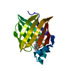
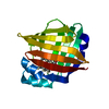
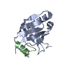
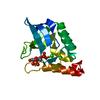
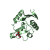
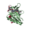
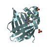
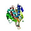
| Title | Enantiomer-Specific Binding of the Potent Antinociceptive Agent SBFI-26 to Anandamide transporters FABP7 | ||||||
 Components Components | Fatty acid-binding protein, brain | ||||||
 Keywords Keywords | LIPID BINDING PROTEIN/INHIBITOR /  inhibitor / LIPID BINDING PROTEIN-INHIBITOR complex inhibitor / LIPID BINDING PROTEIN-INHIBITOR complex | ||||||
| Function / homology |  Function and homology information Function and homology informationNOTCH3 Intracellular Domain Regulates Transcription / Triglyceride catabolism / fatty acid transport / epithelial cell proliferation / fatty acid binding / nervous system development / negative regulation of cell population proliferation / lipid binding / nucleus / cytosolSimilarity search - Function | ||||||
| Biological species |  Homo sapiens (human) Homo sapiens (human) | ||||||
| Method | X-RAY DIFFRACTION / SYNCHROTRON / MOLECULAR REPLACEMENT / Resolution: 1.85002167328 Å | ||||||
 Authors Authors | Hsu, H.-C. / Li, H. | ||||||
| Funding support |  United States, 1items United States, 1items
| ||||||
 Citation Citation | Journal: Biochemistry / Year: 2017 Title: The Antinociceptive Agent SBFI-26 Binds to Anandamide Transporters FABP5 and FABP7 at Two Different Sites. Authors: Hsu, H.C. / Tong, S. / Zhou, Y. / Elmes, M.W. / Yan, S. / Kaczocha, M. / Deutsch, D.G. / Rizzo, R.C. / Ojima, I. / Li, H. | ||||||
| History |
|
- Structure visualization
| Structure viewer | Molecule: MolmilJmol/JSmol |
|---|
- Downloads & links
-Download
| PDBx/mmCIF format | 5ura.cif.gz | 277.3 KB | Display | PDBx/mmCIF format |
|---|---|---|---|---|
| PDB format | pdb5ura.ent.gz | 184.2 KB | Display | PDB format |
| PDBx/mmJSON format | 5ura.json.gz | Tree view | PDBx/mmJSON format | |
| Others |  Other downloads Other downloads |
-Validation report
| Arichive directory | https://data.pdbj.org/pub/pdb/validation_reports/ur/5uraftp://data.pdbj.org/pub/pdb/validation_reports/ur/5ura | HTTPS FTP |
|---|
-Related structure data
| Related structure data |  5ur9C  1fdqS C: citing same article ( S: Starting model for refinement |
|---|---|
| Similar structure data |


-Links
 PDBj
PDBj


- Assembly
| Deposited unit | 
| ||||||||||||
|---|---|---|---|---|---|---|---|---|---|---|---|---|---|
| 1 | 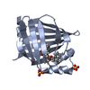
| ||||||||||||
| 2 | 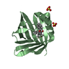
| ||||||||||||
| 3 | 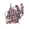
| ||||||||||||
| 4 | 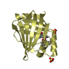
| ||||||||||||
| Unit cell |
| ||||||||||||
| Details | Monomer as determined by gel filtration |
-Components
| #1: Protein | Mass: 15190.174 Da / Num. of mol.: 4 Source method: isolated from a genetically manipulated source Source: (gene. exp.) Homo sapiens (human) / Gene: FABP7, BLBP, FABPB, MRG / Production host:  Escherichia coli (E. coli) / References: UniProt: O15540 Escherichia coli (E. coli) / References: UniProt: O15540#2: Chemical | ChemComp-8KS / (   Mass: 422.472 Da / Num. of mol.: 4 / Source method: obtained synthetically / Formula: C28H22O4 Mass: 422.472 Da / Num. of mol.: 4 / Source method: obtained synthetically / Formula: C28H22O4#3: Chemical | ChemComp-SO4 / Sulfate  Mass: 96.063 Da / Num. of mol.: 8 / Source method: obtained synthetically / Formula: SO4 Mass: 96.063 Da / Num. of mol.: 8 / Source method: obtained synthetically / Formula: SO4#4: Water | ChemComp-HOH / | Water Mass: 18.015 Da / Num. of mol.: 579 / Source method: isolated from a natural source / Formula: H2O Mass: 18.015 Da / Num. of mol.: 579 / Source method: isolated from a natural source / Formula: H2O |
|---|
-Experimental details
-Experiment
| Experiment | Method: X-RAY DIFFRACTION / Number of used crystals: 1 |
|---|
- Sample preparation
| Crystal | Density Matthews: 2.29 Å3/Da / Density % sol: 46.4 % |
|---|---|
| Crystal grow | Temperature: 293 K / Method: vapor diffusion, sitting drop / pH: 8.5 Details: 0.1 M Tris, pH8.5, 0.1 M lithium sulfate, and 33% polyethylene glycol 4000 |
-Data collection
| Diffraction | Mean temperature: 100 K |
|---|---|
| Diffraction source | Source: SYNCHROTRON / Site: APS / Beamline: 17-ID / Wavelength: 0.97931 Å |
| Detector | Type: RAYONIX MX-225 / Detector: CCD / Date: Nov 12, 2015 |
| Radiation | Protocol: SINGLE WAVELENGTH / Monochromatic (M) / Laue (L): M / Scattering type: x-ray |
| Radiation wavelength | Wavelength: 0.97931 Å / Relative weight: 1 |
| Reflection | Resolution: 1.85→53.77 Å / Num. all: 174183 / Num. obs: 46797 / Biso Wilson estimate: 21.2914842067 Å2 / Rmerge(I) obs: 0.107 |
| Reflection shell | Resolution: 1.85→1.95 Å / Redundancy: 3.7 % / Rmerge(I) obs: 0.551 / Mean I/σ(I) obs: 2.3 / Num. unique obs: 6795 / CC1/2: 0.636 / % possible all: 100 |
- Processing
| Software |
| ||||||||||||||||||||||||||||||||||||||||||||||||||||||||||||||||||||||||||||||||||||||||||||||||||||||||||||||||||||||||||||||
|---|---|---|---|---|---|---|---|---|---|---|---|---|---|---|---|---|---|---|---|---|---|---|---|---|---|---|---|---|---|---|---|---|---|---|---|---|---|---|---|---|---|---|---|---|---|---|---|---|---|---|---|---|---|---|---|---|---|---|---|---|---|---|---|---|---|---|---|---|---|---|---|---|---|---|---|---|---|---|---|---|---|---|---|---|---|---|---|---|---|---|---|---|---|---|---|---|---|---|---|---|---|---|---|---|---|---|---|---|---|---|---|---|---|---|---|---|---|---|---|---|---|---|---|---|---|---|---|
| Refinement | Method to determine structure: MOLECULAR REPLACEMENT Starting model: 1FDQ Resolution: 1.85002167328→36.2521887721 Å / SU ML: 0.221034257719 / Cross valid method: FREE R-VALUE / σ(F): 1.35214011819 / Phase error: 22.3950668524 Stereochemistry target values: GeoStd + Monomer Library + CDL v1.2
| ||||||||||||||||||||||||||||||||||||||||||||||||||||||||||||||||||||||||||||||||||||||||||||||||||||||||||||||||||||||||||||||
| Solvent computation | Shrinkage radii: 0.9 Å / VDW probe radii: 1.11 Å / Solvent model: FLAT BULK SOLVENT MODEL | ||||||||||||||||||||||||||||||||||||||||||||||||||||||||||||||||||||||||||||||||||||||||||||||||||||||||||||||||||||||||||||||
| Displacement parameters | Biso mean: 29.9522072011 Å2 | ||||||||||||||||||||||||||||||||||||||||||||||||||||||||||||||||||||||||||||||||||||||||||||||||||||||||||||||||||||||||||||||
| Refinement step | Cycle: LAST / Resolution: 1.85002167328→36.2521887721 Å
| ||||||||||||||||||||||||||||||||||||||||||||||||||||||||||||||||||||||||||||||||||||||||||||||||||||||||||||||||||||||||||||||
| Refine LS restraints |
| ||||||||||||||||||||||||||||||||||||||||||||||||||||||||||||||||||||||||||||||||||||||||||||||||||||||||||||||||||||||||||||||
| LS refinement shell |
|
