 Movie
Movie Controller

[English] 日本語
 Yorodumi
- PDB-5udg: Mutant E97Q crystal structure of Bacillus subtilis QueF with a di... -
+ Open data

- Basic information
| Entry | Database: PDB / ID: 5udg | ||||||
|---|---|---|---|---|---|---|---|
| Title | Mutant E97Q crystal structure of Bacillus subtilis QueF with a disulfide Cys 55-99 | ||||||
 Components Components | NADPH-dependent 7-cyano-7-deazaguanine reductase | ||||||
 Keywords Keywords |  OXIDOREDUCTASE / Tunnel fold / Disulfide inactivation / tRNA modification pathway / NADPH-dependent reduction of the nitrile group OXIDOREDUCTASE / Tunnel fold / Disulfide inactivation / tRNA modification pathway / NADPH-dependent reduction of the nitrile group | ||||||
| Function / homology |  Function and homology informationpreQ1 synthase / preQ1 synthase activity / oxidoreductase activity, acting on other nitrogenous compounds as donors, with NAD or NADP as acceptor / queuosine biosynthetic process / metal ion binding / cytoplasm Function and homology informationpreQ1 synthase / preQ1 synthase activity / oxidoreductase activity, acting on other nitrogenous compounds as donors, with NAD or NADP as acceptor / queuosine biosynthetic process / metal ion binding / cytoplasmSimilarity search - Function | ||||||
| Biological species |  Bacillus subtilis (bacteria) Bacillus subtilis (bacteria) | ||||||
| Method | X-RAY DIFFRACTION / SYNCHROTRON / MOLECULAR REPLACEMENT / Resolution: 2.5 Å | ||||||
 Authors Authors | Mohammad, A. / Kiani, M.K. / Iwata-Reuyl, D. / Stec, B. / Swairjo, M. | ||||||
 Citation Citation | Journal: Biomolecules / Year: 2017 Title: Protection of the Queuosine Biosynthesis Enzyme QueF from Irreversible Oxidation by a Conserved Intramolecular Disulfide. Authors: Mohammad, A. / Bon Ramos, A. / Lee, B.W. / Cohen, S.W. / Kiani, M.K. / Iwata-Reuyl, D. / Stec, B. / Swairjo, M.A. | ||||||
| History |
|
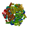
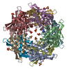
- Structure visualization
| Structure viewer | Molecule: MolmilJmol/JSmol |
|---|
- Downloads & links
-Download
| PDBx/mmCIF format | 5udg.cif.gz | 170.9 KB | Display | PDBx/mmCIF format |
|---|---|---|---|---|
| PDB format | pdb5udg.ent.gz | 135.2 KB | Display | PDB format |
| PDBx/mmJSON format | 5udg.json.gz | Tree view | PDBx/mmJSON format | |
| Others |  Other downloads Other downloads |
-Validation report
| Arichive directory | https://data.pdbj.org/pub/pdb/validation_reports/ud/5udgftp://data.pdbj.org/pub/pdb/validation_reports/ud/5udg | HTTPS FTP |
|---|
-Related structure data
| Related structure data | 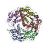 4f8bS S: Starting model for refinement |
|---|---|
| Similar structure data |


-Links
 PDBj
PDBj
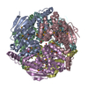
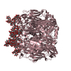
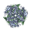
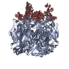
- Assembly
| Deposited unit | 
| ||||||||
|---|---|---|---|---|---|---|---|---|---|
| 1 | 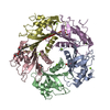
| ||||||||
| Unit cell |
|
-Components
| #1: Protein | Mass: 17247.570 Da / Num. of mol.: 5 / Fragment: UNP resdiues 21-165 / Mutation: E97Q Source method: isolated from a genetically manipulated source Source: (gene. exp.) Bacillus subtilis (bacteria)Gene: queF, A9D36_18900, AX282_02560, B4122_4589, B4417_3799, BN2127_JRS11_03356, BN2127_JRS2_02117, BN2127_JRS6_01198, BN2127_JRS9_01727, SC09_Contig19orf00807 Production host: Escherichia coli (E. coli)References: UniProt: A0A063X9I2, UniProt: O31678*PLUS, preQ1 synthase#2: Chemical | ChemComp-MG /   Mass: 24.305 Da / Num. of mol.: 7 / Source method: obtained synthetically / Formula: Mg Mass: 24.305 Da / Num. of mol.: 7 / Source method: obtained synthetically / Formula: Mg#3: Chemical | ChemComp-PGE / Polyethylene glycol  Mass: 150.173 Da / Num. of mol.: 4 / Source method: obtained synthetically / Formula: C6H14O4 Mass: 150.173 Da / Num. of mol.: 4 / Source method: obtained synthetically / Formula: C6H14O4#4: Water | ChemComp-HOH / | Water Mass: 18.015 Da / Num. of mol.: 285 / Source method: isolated from a natural source / Formula: H2O Mass: 18.015 Da / Num. of mol.: 285 / Source method: isolated from a natural source / Formula: H2O |
|---|
-Experimental details
-Experiment
| Experiment | Method: X-RAY DIFFRACTION / Number of used crystals: 1 |
|---|
- Sample preparation
| Crystal | Density Matthews: 2.51 Å3/Da / Density % sol: 51 % / Description: Rhomboidal |
|---|---|
| Crystal grow | Temperature: 293 K / Method: vapor diffusion, hanging drop Details: Protein sample 4 mg/mL, in 50 mM Tris pH 7.5, 50 mM KCl, 1 mM MgCl2 and 1 mM (mercaptoethanol-BME) was mixed with reservoir solution (1:1 ratio) containing 19% PEG 550 monomethyl ether, 43 ...Details: Protein sample 4 mg/mL, in 50 mM Tris pH 7.5, 50 mM KCl, 1 mM MgCl2 and 1 mM (mercaptoethanol-BME) was mixed with reservoir solution (1:1 ratio) containing 19% PEG 550 monomethyl ether, 43 mM imidazole-Cl (pH 6.8), 53 mM imidazole (pH 8.2), 30 mM CaCl2, and 4% DMSO |
-Data collection
| Diffraction | Mean temperature: 100 K |
|---|---|
| Diffraction source | Source: SYNCHROTRON / Site: SSRL  / Beamline: BL12-2 / Wavelength: 1.1 Å / Beamline: BL12-2 / Wavelength: 1.1 Å |
| Detector | Type: MARRESEARCH / Detector: CCD / Date: Dec 12, 2012 |
| Radiation | Protocol: SINGLE WAVELENGTH / Monochromatic (M) / Laue (L): M / Scattering type: x-ray |
| Radiation wavelength | Wavelength: 1.1 Å / Relative weight: 1 |
| Reflection | Resolution: 2.5→45.61 Å / Num. obs: 30298 / % possible obs: 98 % / Observed criterion σ(F): 0 / Observed criterion σ(I): 0 / Redundancy: 5 % / Biso Wilson estimate: 50 Å2 / Rmerge(I) obs: 0.121 / Net I/σ(I): 14 |
| Reflection shell | Resolution: 2.5→2.54 Å / Redundancy: 3 % / Rmerge(I) obs: 0.83 / CC1/2: 0.75 / % possible all: 89.4 |
- Processing
| Software |
| ||||||||||||||||||||||||||||||||||||||||||||||||||||||||||||||||||||||||||||||||||||||||||||||||||||||||||||||||||||||||||||||||||||||||||||||||||||||||||||||||||||||||||||||||||||||
|---|---|---|---|---|---|---|---|---|---|---|---|---|---|---|---|---|---|---|---|---|---|---|---|---|---|---|---|---|---|---|---|---|---|---|---|---|---|---|---|---|---|---|---|---|---|---|---|---|---|---|---|---|---|---|---|---|---|---|---|---|---|---|---|---|---|---|---|---|---|---|---|---|---|---|---|---|---|---|---|---|---|---|---|---|---|---|---|---|---|---|---|---|---|---|---|---|---|---|---|---|---|---|---|---|---|---|---|---|---|---|---|---|---|---|---|---|---|---|---|---|---|---|---|---|---|---|---|---|---|---|---|---|---|---|---|---|---|---|---|---|---|---|---|---|---|---|---|---|---|---|---|---|---|---|---|---|---|---|---|---|---|---|---|---|---|---|---|---|---|---|---|---|---|---|---|---|---|---|---|---|---|---|---|
| Refinement | Method to determine structure: MOLECULAR REPLACEMENT Starting model: 4F8B Resolution: 2.5→45.61 Å / Cor.coef. Fo:Fc: 0.949 / Cor.coef. Fo:Fc free: 0.89 / SU B: 12.067 / SU ML: 0.265 / Cross valid method: THROUGHOUT / ESU R: 0.723 / ESU R Free: 0.355 / Details: HYDROGENS HAVE BEEN USED IF PRESENT IN THE INPUT
| ||||||||||||||||||||||||||||||||||||||||||||||||||||||||||||||||||||||||||||||||||||||||||||||||||||||||||||||||||||||||||||||||||||||||||||||||||||||||||||||||||||||||||||||||||||||
| Solvent computation | Ion probe radii: 0.8 Å / Shrinkage radii: 0.8 Å / VDW probe radii: 1.2 Å | ||||||||||||||||||||||||||||||||||||||||||||||||||||||||||||||||||||||||||||||||||||||||||||||||||||||||||||||||||||||||||||||||||||||||||||||||||||||||||||||||||||||||||||||||||||||
| Displacement parameters | Biso mean: 47.938 Å2
| ||||||||||||||||||||||||||||||||||||||||||||||||||||||||||||||||||||||||||||||||||||||||||||||||||||||||||||||||||||||||||||||||||||||||||||||||||||||||||||||||||||||||||||||||||||||
| Refinement step | Cycle: 1 / Resolution: 2.5→45.61 Å
| ||||||||||||||||||||||||||||||||||||||||||||||||||||||||||||||||||||||||||||||||||||||||||||||||||||||||||||||||||||||||||||||||||||||||||||||||||||||||||||||||||||||||||||||||||||||
| Refine LS restraints |
|