 Movie
Movie Controller


+ Open data

- Basic information
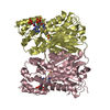
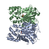
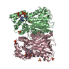
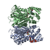
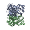
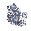
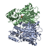
| Entry | Database: PDB / ID: 5uco | ||||||
|---|---|---|---|---|---|---|---|
| Title | Benzophenone synthase from Hypericum androsaemum | ||||||
 Components Components | 2,4,6-trihydroxybenzophenone synthase | ||||||
 Keywords Keywords |  TRANSFERASE / thiolase / polyketide / benzoyl-CoA TRANSFERASE / thiolase / polyketide / benzoyl-CoA | ||||||
| Function / homology |  Function and homology information Function and homology information2,3',4,6-tetrahydroxybenzophenone synthase / 2,4,6-trihydroxybenzophenone synthase / tetrahydroxybenzophenone synthase activity / trihydroxybenzophenone synthase activity / benzoyl-CoA metabolic process / malonyl-CoA metabolic process / biosynthetic process / acyltransferase activitySimilarity search - Function | ||||||
| Biological species |  Hypericum androsaemum (plant) Hypericum androsaemum (plant) | ||||||
| Method | X-RAY DIFFRACTION / SYNCHROTRON / MOLECULAR REPLACEMENT / molecular replacement / Resolution: 2.85 Å | ||||||
 Authors Authors | Stewart Jr, C.E. / Noel, J.P. | ||||||
 Citation Citation | Journal: Acta Crystallogr D Struct Biol / Year: 2017 Title: Molecular architectures of benzoic acid-specific type III polyketide synthases. Authors: Stewart, C. / Woods, K. / Macias, G. / Allan, A.C. / Hellens, R.P. / Noel, J.P. | ||||||
| History |
|
- Structure visualization
| Structure viewer | Molecule: MolmilJmol/JSmol |
|---|
- Downloads & links
-Download
| PDBx/mmCIF format | 5uco.cif.gz | 271.4 KB | Display | PDBx/mmCIF format |
|---|---|---|---|---|
| PDB format | pdb5uco.ent.gz | 220.8 KB | Display | PDB format |
| PDBx/mmJSON format | 5uco.json.gz | Tree view | PDBx/mmJSON format | |
| Others |  Other downloads Other downloads |
-Validation report
| Arichive directory | https://data.pdbj.org/pub/pdb/validation_reports/uc/5ucoftp://data.pdbj.org/pub/pdb/validation_reports/uc/5uco | HTTPS FTP |
|---|
-Related structure data
| Related structure data |  5uc5C  5w8qC  5wc4C 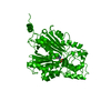 1bi5S S: Starting model for refinement C: citing same article ( |
|---|---|
| Similar structure data |


-Links
 PDBj
PDBj

- Assembly
| Deposited unit | 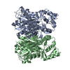
| ||||||||
|---|---|---|---|---|---|---|---|---|---|
| 1 |
| ||||||||
| Unit cell |
|
-Components
| #1: Protein | Mass: 43017.988 Da / Num. of mol.: 2 Source method: isolated from a genetically manipulated source Details: codon optimized for E.coli expression, His-tag removed after purification Source: (gene. exp.) Hypericum androsaemum (plant) / Gene: BPS / Plasmid: pHIS8 / Production host:  Escherichia coli (E. coli) / Strain (production host): BL21(DE3) Escherichia coli (E. coli) / Strain (production host): BL21(DE3)References: UniProt: Q8SAS8, 2,3',4,6-tetrahydroxybenzophenone synthase #2: Water | ChemComp-HOH / | Water Mass: 18.015 Da / Num. of mol.: 44 / Source method: isolated from a natural source / Formula: H2O Mass: 18.015 Da / Num. of mol.: 44 / Source method: isolated from a natural source / Formula: H2O |
|---|
-Experimental details
-Experiment
| Experiment | Method: X-RAY DIFFRACTION / Number of used crystals: 1 |
|---|
- Sample preparation
| Crystal | Density Matthews: 2.72 Å3/Da / Density % sol: 54.79 % |
|---|---|
| Crystal grow | Temperature: 278 K / Method: vapor diffusion, hanging drop / pH: 6.5 Details: Reservoir: PIPES (pH 6.5) 100mM, PEG8000 18%; ratio of protein to reservoir = 1:1 |
-Data collection
| Diffraction | Mean temperature: 100 K | ||||||||||||||||||||||||||||||||||||||||||||||||||||||||||||||||||
|---|---|---|---|---|---|---|---|---|---|---|---|---|---|---|---|---|---|---|---|---|---|---|---|---|---|---|---|---|---|---|---|---|---|---|---|---|---|---|---|---|---|---|---|---|---|---|---|---|---|---|---|---|---|---|---|---|---|---|---|---|---|---|---|---|---|---|---|
| Diffraction source | Source: SYNCHROTRON / Site: ALS  / Beamline: 8.2.1 / Wavelength: 1 Å / Beamline: 8.2.1 / Wavelength: 1 Å | ||||||||||||||||||||||||||||||||||||||||||||||||||||||||||||||||||
| Detector | Type: ADSC QUANTUM 315r / Detector: CCD / Date: Apr 5, 2013 / Details: mirrors | ||||||||||||||||||||||||||||||||||||||||||||||||||||||||||||||||||
| Radiation | Monochromator: double crystal Si(111) / Protocol: SINGLE WAVELENGTH / Scattering type: x-ray | ||||||||||||||||||||||||||||||||||||||||||||||||||||||||||||||||||
| Radiation wavelength | Wavelength: 1 Å / Relative weight: 1 | ||||||||||||||||||||||||||||||||||||||||||||||||||||||||||||||||||
| Reflection | Resolution: 2.85→184.97 Å / Num. all: 18840 / Num. obs: 18840 / % possible obs: 85 % / Redundancy: 3.5 % / Biso Wilson estimate: 21.7 Å2 / Rpim(I) all: 0.142 / Rrim(I) all: 0.294 / Rsym value: 0.253 / Net I/av σ(I): 2.2 / Net I/σ(I): 4.5 / Num. measured all: 65535 | ||||||||||||||||||||||||||||||||||||||||||||||||||||||||||||||||||
| Reflection shell |
|
-Phasing
| Phasing | Method: molecular replacement |
|---|
- Processing
| Software |
| ||||||||||||||||||||||||||||||||||||||||||||||||||||||||||||||||||||||||||||||||||||||||||||||||||
|---|---|---|---|---|---|---|---|---|---|---|---|---|---|---|---|---|---|---|---|---|---|---|---|---|---|---|---|---|---|---|---|---|---|---|---|---|---|---|---|---|---|---|---|---|---|---|---|---|---|---|---|---|---|---|---|---|---|---|---|---|---|---|---|---|---|---|---|---|---|---|---|---|---|---|---|---|---|---|---|---|---|---|---|---|---|---|---|---|---|---|---|---|---|---|---|---|---|---|---|
| Refinement | Method to determine structure: MOLECULAR REPLACEMENT Starting model: 1BI5 Resolution: 2.85→46.242 Å / SU ML: 0.37 / Cross valid method: FREE R-VALUE / σ(F): 1.35 / Phase error: 24.09
| ||||||||||||||||||||||||||||||||||||||||||||||||||||||||||||||||||||||||||||||||||||||||||||||||||
| Solvent computation | Shrinkage radii: 0.9 Å / VDW probe radii: 1.11 Å | ||||||||||||||||||||||||||||||||||||||||||||||||||||||||||||||||||||||||||||||||||||||||||||||||||
| Displacement parameters | Biso max: 55.38 Å2 / Biso mean: 15.6359 Å2 / Biso min: 2.58 Å2 | ||||||||||||||||||||||||||||||||||||||||||||||||||||||||||||||||||||||||||||||||||||||||||||||||||
| Refinement step | Cycle: final / Resolution: 2.85→46.242 Å
| ||||||||||||||||||||||||||||||||||||||||||||||||||||||||||||||||||||||||||||||||||||||||||||||||||
| Refine LS restraints |
| ||||||||||||||||||||||||||||||||||||||||||||||||||||||||||||||||||||||||||||||||||||||||||||||||||
| LS refinement shell | Refine-ID: X-RAY DIFFRACTION / Rfactor Rfree error: 0 / Total num. of bins used: 13
|
