 Movie
Movie Controller


+ Open data

- Basic information
| Entry | Database: PDB / ID: 5ubw | ||||||
|---|---|---|---|---|---|---|---|
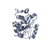
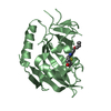
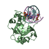
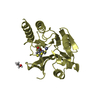
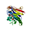
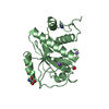
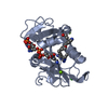
| Title | Structure of catalytic domain of Ssel | ||||||
 Components Components | Deubiquitinase SseL | ||||||
 Keywords Keywords |  HYDROLASE / UCH family / Salmonella / deubiquitinases HYDROLASE / UCH family / Salmonella / deubiquitinases | ||||||
| Function / homology | cysteine-type peptidase activity / host cell cytoplasm / Hydrolases; Acting on peptide bonds (peptidases); Cysteine endopeptidases / proteolysis / extracellular region / Deubiquitinase SseL Function and homology information Function and homology information | ||||||
| Biological species |  Salmonella typhimurium (bacteria) Salmonella typhimurium (bacteria) | ||||||
| Method | X-RAY DIFFRACTION / SYNCHROTRON / MOLECULAR REPLACEMENT / Resolution: 2.394 Å | ||||||
 Authors Authors | Shrestha, R. / Das, C. | ||||||
| Funding support |  United States, 1items United States, 1items
| ||||||
 Citation Citation | Journal: To Be Published Title: Structure of catalytic domain of Ssel Authors: Shrestha, R. / Das, C. | ||||||
| History |
|
- Structure visualization
| Structure viewer | Molecule: MolmilJmol/JSmol |
|---|
- Downloads & links
-Download
| PDBx/mmCIF format | 5ubw.cif.gz | 81.2 KB | Display | PDBx/mmCIF format |
|---|---|---|---|---|
| PDB format | pdb5ubw.ent.gz | 59.3 KB | Display | PDB format |
| PDBx/mmJSON format | 5ubw.json.gz | Tree view | PDBx/mmJSON format | |
| Others |  Other downloads Other downloads |
-Validation report
| Arichive directory | https://data.pdbj.org/pub/pdb/validation_reports/ub/5ubwftp://data.pdbj.org/pub/pdb/validation_reports/ub/5ubw | HTTPS FTP |
|---|
-Related structure data
| Related structure data |  5hafS S: Starting model for refinement |
|---|---|
| Similar structure data |
-Links
 PDBj
PDBj- Assembly
| Deposited unit | 
| ||||||||
|---|---|---|---|---|---|---|---|---|---|
| 1 | 
| ||||||||
| 2 | 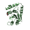
| ||||||||
| Unit cell |
|
-Components
| #1: Protein | Mass: 21033.674 Da / Num. of mol.: 2 Source method: isolated from a genetically manipulated source Source: (gene. exp.) Salmonella typhimurium (bacteria) / Strain: LT2 / SGSC1412 / ATCC 700720 / Gene: sseL, STM2287 / Production host: Escherichia coli (E. coli)References: UniProt: Q8ZNG2, Hydrolases; Acting on peptide bonds (peptidases); Cysteine endopeptidases#2: Chemical | ChemComp-EDO / | Ethylene glycol  Mass: 62.068 Da / Num. of mol.: 1 / Source method: obtained synthetically / Formula: C2H6O2 Mass: 62.068 Da / Num. of mol.: 1 / Source method: obtained synthetically / Formula: C2H6O2#3: Water | ChemComp-HOH / | Water Mass: 18.015 Da / Num. of mol.: 22 / Source method: isolated from a natural source / Formula: H2O Mass: 18.015 Da / Num. of mol.: 22 / Source method: isolated from a natural source / Formula: H2O |
|---|
-Experimental details
-Experiment
| Experiment | Method: X-RAY DIFFRACTION / Number of used crystals: 1 |
|---|
- Sample preparation
| Crystal | Density Matthews: 2.35 Å3/Da / Density % sol: 47.77 % |
|---|---|
| Crystal grow | Temperature: 295 K / Method: vapor diffusion / pH: 7.5 Details: 0.1 M HEPES sodium pH 7.5 and 1.5 M lithium sulfate monohydrate |
-Data collection
| Diffraction | Mean temperature: 100 K |
|---|---|
| Diffraction source | Source: SYNCHROTRON / Site: APS / Beamline: 23-ID-B / Wavelength: 1 Å |
| Detector | Type: MAR scanner 300 mm plate / Detector: IMAGE PLATE / Date: Aug 9, 2015 |
| Radiation | Protocol: SINGLE WAVELENGTH / Monochromatic (M) / Laue (L): M / Scattering type: x-ray |
| Radiation wavelength | Wavelength: 1 Å / Relative weight: 1 |
| Reflection | Resolution: 2.394→54.6 Å / Num. obs: 14585 / % possible obs: 99.9 % / Redundancy: 3.8 % / Net I/σ(I): 11.4 |
| Reflection shell | Resolution: 2.394→2.44 Å / Redundancy: 3.8 % / % possible all: 100 |
- Processing
| Software |
| ||||||||||||||||||||||||||||||||||||||||||
|---|---|---|---|---|---|---|---|---|---|---|---|---|---|---|---|---|---|---|---|---|---|---|---|---|---|---|---|---|---|---|---|---|---|---|---|---|---|---|---|---|---|---|---|
| Refinement | Method to determine structure: MOLECULAR REPLACEMENT Starting model: 5HAF Resolution: 2.394→42.604 Å / SU ML: 0.4 / Cross valid method: THROUGHOUT / σ(F): 1.38 / Phase error: 34.88
| ||||||||||||||||||||||||||||||||||||||||||
| Solvent computation | Shrinkage radii: 0.9 Å / VDW probe radii: 1.11 Å | ||||||||||||||||||||||||||||||||||||||||||
| Refinement step | Cycle: LAST / Resolution: 2.394→42.604 Å
| ||||||||||||||||||||||||||||||||||||||||||
| Refine LS restraints |
| ||||||||||||||||||||||||||||||||||||||||||
| LS refinement shell |
|
