 Movie
Movie Controller

[English] 日本語
 Yorodumi
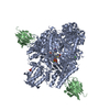
- PDB-5u68: Structural basis for antibody cross-neutralization of respiratory... -
+ Open data

- Basic information
| Entry | Database: PDB / ID: 5u68 | ||||||
|---|---|---|---|---|---|---|---|
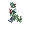
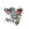
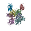
| Title | Structural basis for antibody cross-neutralization of respiratory syncytial virus and human metapneumovirus | ||||||
 Components Components |
| ||||||
 Keywords Keywords |  VIRAL PROTEIN/Immune System / RSVF prefusion / MPE8 / cross-reactive antibody neutralization / Trimer / VIRAL PROTEIN-Immune System complex VIRAL PROTEIN/Immune System / RSVF prefusion / MPE8 / cross-reactive antibody neutralization / Trimer / VIRAL PROTEIN-Immune System complex | ||||||
| Function / homology |  Function and homology information Function and homology informationpositive regulation of syncytium formation by virus / host cell Golgi membrane / entry receptor-mediated virion attachment to host cell / symbiont entry into host cell / fusion of virus membrane with host plasma membrane / viral envelope / host cell plasma membrane / virion membrane / membrane / identical protein bindingSimilarity search - Function | ||||||
| Biological species |  Human respiratory syncytial virus A Human respiratory syncytial virus A Human immunodeficiency virus 1 Human immunodeficiency virus 1 Homo sapiens (human) Homo sapiens (human) | ||||||
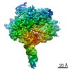
| Method | X-RAY DIFFRACTION / SYNCHROTRON / MOLECULAR REPLACEMENT / Resolution: 3.083 Å | ||||||
 Authors Authors | Wen, X. / Jardetzky, T.S. | ||||||
| Funding support |  United States, 1items United States, 1items
| ||||||
 Citation Citation | Journal: Nat Microbiol / Year: 2017 Title: Structural basis for antibody cross-neutralization of respiratory syncytial virus and human metapneumovirus. Authors: Wen, X. / Mousa, J.J. / Bates, J.T. / Lamb, R.A. / Crowe, J.E. / Jardetzky, T.S. | ||||||
| History |
|
- Structure visualization
| Structure viewer | Molecule: MolmilJmol/JSmol |
|---|
- Downloads & links
-Download
| PDBx/mmCIF format | 5u68.cif.gz | 1.1 MB | Display | PDBx/mmCIF format |
|---|---|---|---|---|
| PDB format | pdb5u68.ent.gz | 965.4 KB | Display | PDB format |
| PDBx/mmJSON format | 5u68.json.gz | Tree view | PDBx/mmJSON format | |
| Others |  Other downloads Other downloads |
-Validation report
| Arichive directory | https://data.pdbj.org/pub/pdb/validation_reports/u6/5u68ftp://data.pdbj.org/pub/pdb/validation_reports/u6/5u68 | HTTPS FTP |
|---|
-Related structure data


-Links
 PDBj
PDBj


- Assembly
| Deposited unit | 
| ||||||||
|---|---|---|---|---|---|---|---|---|---|
| 1 |
| ||||||||
| Unit cell |
|
-Components
| #1: Protein | / Protein F Mass: 62452.488 Da / Num. of mol.: 3 Fragment: UNP P03420 residues 1-513,UNP M1E1E4 residues 1-28 Source method: isolated from a genetically manipulated source Source: (gene. exp.) Human respiratory syncytial virus A (strain A2), (gene. exp.) Human immunodeficiency virus 1Strain: A2 / Plasmid: pTT5 / Cell line (production host): 293-6E / Production host: Homo sapiens (human) / References: UniProt: P03420, UniProt: M1E1E4#2: Antibody | Mass: 30684.664 Da / Num. of mol.: 3 Source method: isolated from a genetically manipulated source Details: single chain antibody (scFv) / Source: (gene. exp.) Homo sapiens (human) / Production host: Homo sapiens (human) |
|---|
-Experimental details
-Experiment
| Experiment | Method: X-RAY DIFFRACTION / Number of used crystals: 1 |
|---|
- Sample preparation
| Crystal | Density Matthews: 3.45 Å3/Da / Density % sol: 64.36 % / Description: rod-shaped |
|---|---|
| Crystal grow | Temperature: 295 K / Method: vapor diffusion, hanging drop / pH: 7 Details: 0.1M Potassium Nitrate, 0.1M Citrate Phosphate pH 4.2, 1 % Tacsimate pH 7.0, 14 (w/v) % PEG 6000 PH range: 7.0-7.4 |
-Data collection
| Diffraction | Mean temperature: 100 K |
|---|---|
| Diffraction source | Source: SYNCHROTRON / Site: APS / Beamline: 21-ID-F / Wavelength: 0.97872 Å |
| Detector | Type: RAYONIX MX-225 / Detector: CCD / Date: Aug 13, 2015 / Details: Fibre Optic Taper (FOT) |
| Radiation | Monochromator: C(111) / Protocol: SINGLE WAVELENGTH / Monochromatic (M) / Laue (L): M / Scattering type: x-ray |
| Radiation wavelength | Wavelength: 0.97872 Å / Relative weight: 1 |
| Reflection | Resolution: 3.1→48.191 Å / Num. obs: 71891 / % possible obs: 99.98 % / Redundancy: 11.2 % / Net I/σ(I): 11.96 |
- Processing
| Software |
| ||||||||||||||||||||||||||||||||||||||||||||||||||||||||||||||||||||||||||||||||||||||||||||||||||||||||||||||||||||||||||||||||||||||||||||
|---|---|---|---|---|---|---|---|---|---|---|---|---|---|---|---|---|---|---|---|---|---|---|---|---|---|---|---|---|---|---|---|---|---|---|---|---|---|---|---|---|---|---|---|---|---|---|---|---|---|---|---|---|---|---|---|---|---|---|---|---|---|---|---|---|---|---|---|---|---|---|---|---|---|---|---|---|---|---|---|---|---|---|---|---|---|---|---|---|---|---|---|---|---|---|---|---|---|---|---|---|---|---|---|---|---|---|---|---|---|---|---|---|---|---|---|---|---|---|---|---|---|---|---|---|---|---|---|---|---|---|---|---|---|---|---|---|---|---|---|---|---|
| Refinement | Method to determine structure: MOLECULAR REPLACEMENT Starting model: 4JHW, 3H42 Resolution: 3.083→48.191 Å / SU ML: 0.36 / Cross valid method: FREE R-VALUE / σ(F): 1.36 / Phase error: 23.9 / Stereochemistry target values: ML
| ||||||||||||||||||||||||||||||||||||||||||||||||||||||||||||||||||||||||||||||||||||||||||||||||||||||||||||||||||||||||||||||||||||||||||||
| Solvent computation | Shrinkage radii: 0.9 Å / VDW probe radii: 1.11 Å / Solvent model: FLAT BULK SOLVENT MODEL | ||||||||||||||||||||||||||||||||||||||||||||||||||||||||||||||||||||||||||||||||||||||||||||||||||||||||||||||||||||||||||||||||||||||||||||
| Refinement step | Cycle: LAST / Resolution: 3.083→48.191 Å
| ||||||||||||||||||||||||||||||||||||||||||||||||||||||||||||||||||||||||||||||||||||||||||||||||||||||||||||||||||||||||||||||||||||||||||||
| Refine LS restraints |
| ||||||||||||||||||||||||||||||||||||||||||||||||||||||||||||||||||||||||||||||||||||||||||||||||||||||||||||||||||||||||||||||||||||||||||||
| LS refinement shell |
| ||||||||||||||||||||||||||||||||||||||||||||||||||||||||||||||||||||||||||||||||||||||||||||||||||||||||||||||||||||||||||||||||||||||||||||
| Refinement TLS params. | Method: refined / Origin x: -135.4741 Å / Origin y: 162.3403 Å / Origin z: 288.1867 Å
| ||||||||||||||||||||||||||||||||||||||||||||||||||||||||||||||||||||||||||||||||||||||||||||||||||||||||||||||||||||||||||||||||||||||||||||
| Refinement TLS group | Selection details: all |
