 Movie
Movie Controller

[English] 日本語
 Yorodumi
- PDB-5u42: Human PPARdelta ligand-binding domain in complexed with specific ... -
+ Open data

- Basic information
| Entry | Database: PDB / ID: 5u42 | |||||||||
|---|---|---|---|---|---|---|---|---|---|---|
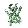
| Title | Human PPARdelta ligand-binding domain in complexed with specific agonist 11 | |||||||||
 Components Components | Peroxisome proliferator-activated receptor delta | |||||||||
 Keywords Keywords | protein binding/activator / PPARdelta / ligand-binding domain / agonist / protein binding-activator complex | |||||||||
| Function / homology |  Function and homology information Function and homology informationfat cell proliferation / positive regulation of fat cell proliferation / keratinocyte migration / linoleic acid binding / positive regulation of skeletal muscle tissue regeneration / axon ensheathment / regulation of skeletal muscle satellite cell proliferation / glucose transmembrane transport / positive regulation of myoblast proliferation / fatty acid catabolic process ...fat cell proliferation / positive regulation of fat cell proliferation / keratinocyte migration / linoleic acid binding / positive regulation of skeletal muscle tissue regeneration / axon ensheathment / regulation of skeletal muscle satellite cell proliferation / glucose transmembrane transport / positive regulation of myoblast proliferation / fatty acid catabolic process / negative regulation of myoblast differentiation / Carnitine metabolism / Regulation of pyruvate dehydrogenase (PDH) complex / Signaling by Retinoic Acid / nuclear steroid receptor activity / fatty acid beta-oxidation / positive regulation of fatty acid metabolic process / cell-substrate adhesion / negative regulation of cholesterol storage / cellular response to nutrient levels / decidualization / keratinocyte proliferation / positive regulation of fat cell differentiation / fatty acid transport / adipose tissue development / energy homeostasis / embryo implantation / cholesterol metabolic process / hormone-mediated signaling pathway / fatty acid metabolic process / negative regulation of miRNA transcription / phosphatidylinositol 3-kinase/protein kinase B signal transduction / generation of precursor metabolites and energy / apoptotic signaling pathway / wound healing / lipid metabolic process / transcription coactivator binding / DNA-binding transcription repressor activity, RNA polymerase II-specific / negative regulation of inflammatory response / Nuclear Receptor transcription pathway / glucose metabolic process / nuclear receptor activity / negative regulation of epithelial cell proliferation / sequence-specific double-stranded DNA binding / cellular response to hypoxia / DNA-binding transcription factor binding / cell population proliferation / cell differentiation / positive regulation of phosphatidylinositol 3-kinase/protein kinase B signal transduction / DNA-binding transcription factor activity, RNA polymerase II-specific / RNA polymerase II cis-regulatory region sequence-specific DNA binding / DNA-binding transcription factor activity / negative regulation of DNA-templated transcription / apoptotic process / lipid binding / chromatin / positive regulation of gene expression / regulation of transcription by RNA polymerase II / positive regulation of DNA-templated transcription / negative regulation of transcription by RNA polymerase II / positive regulation of transcription by RNA polymerase II / DNA binding / zinc ion binding / nucleoplasm / nucleusSimilarity search - Function | |||||||||
| Biological species |  Homo sapiens (human) Homo sapiens (human) | |||||||||
| Method | X-RAY DIFFRACTION / SYNCHROTRON / MOLECULAR REPLACEMENT / Resolution: 1.7 Å | |||||||||
 Authors Authors | Wu, C.-C. / Baiga, T.J. / Downes, M. / La Clair, J.J. / Atkins, A.R. / Richard, S.B. / Stockley-Noel, T.A. / Bowman, M.E. / Evans, R.M. / Noel, J.P. | |||||||||
 Citation Citation | Journal: Proc. Natl. Acad. Sci. U.S.A. / Year: 2017 Title: Structural basis for specific ligation of the peroxisome proliferator-activated receptor delta. Authors: Wu, C.C. / Baiga, T.J. / Downes, M. / La Clair, J.J. / Atkins, A.R. / Richard, S.B. / Fan, W. / Stockley-Noel, T.A. / Bowman, M.E. / Noel, J.P. / Evans, R.M. | |||||||||
| History |
|
- Structure visualization
| Structure viewer | Molecule: MolmilJmol/JSmol |
|---|
- Downloads & links
-Download
| PDBx/mmCIF format | 5u42.cif.gz | 350.6 KB | Display | PDBx/mmCIF format |
|---|---|---|---|---|
| PDB format | pdb5u42.ent.gz | 294.7 KB | Display | PDB format |
| PDBx/mmJSON format | 5u42.json.gz | Tree view | PDBx/mmJSON format | |
| Others |  Other downloads Other downloads |
-Validation report
| Arichive directory | https://data.pdbj.org/pub/pdb/validation_reports/u4/5u42ftp://data.pdbj.org/pub/pdb/validation_reports/u4/5u42 | HTTPS FTP |
|---|
-Related structure data
| Related structure data |  5u3qC  5u3rC  5u3sC  5u3tC  5u3uC  5u3vC  5u3wC  5u3xC  5u3yC 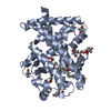 5u3zC 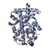 5u40C  5u41C 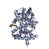 5u43C 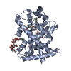 5u44C  5u45C  5u46C  1gwzS C: citing same article ( S: Starting model for refinement |
|---|---|
| Similar structure data |


-Links
 PDBj
PDBj
- Assembly
| Deposited unit | 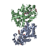
| ||||||||
|---|---|---|---|---|---|---|---|---|---|
| 1 | 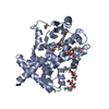
| ||||||||
| 2 | 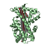
| ||||||||
| Unit cell |
|
-Components
-Protein / Sugars , 2 types, 5 molecules AB
| #1: Protein | / PPAR-delta / NUCI / Nuclear hormone receptor 1 / NUC1 / Nuclear receptor subfamily 1 group C member ...PPAR-delta / NUCI / Nuclear hormone receptor 1 / NUC1 / Nuclear receptor subfamily 1 group C member 2 / Peroxisome proliferator-activated receptor beta / PPAR-beta Mass: 31114.178 Da / Num. of mol.: 2 / Fragment: Ligand-binding domain Source method: isolated from a genetically manipulated source Source: (gene. exp.) Homo sapiens (human) / Gene: PPARD, NR1C2, PPARB / Plasmid: pET28a(+) / Production host:  Escherichia coli BL21(DE3) (bacteria) / References: UniProt: Q03181 Escherichia coli BL21(DE3) (bacteria) / References: UniProt: Q03181#2: Sugar |  Type: D-saccharide / Mass: 278.342 Da / Num. of mol.: 3 / Source method: obtained synthetically / Formula: C13H26O6 Type: D-saccharide / Mass: 278.342 Da / Num. of mol.: 3 / Source method: obtained synthetically / Formula: C13H26O6 |
|---|
-Non-polymers , 4 types, 362 molecules 


| #3: Chemical |  Mass: 487.587 Da / Num. of mol.: 2 / Source method: obtained synthetically / Formula: C30H33NO5 Mass: 487.587 Da / Num. of mol.: 2 / Source method: obtained synthetically / Formula: C30H33NO5#4: Chemical | ChemComp-PGO / Propanediol Mass: 76.094 Da / Num. of mol.: 4 / Source method: obtained synthetically / Formula: C3H8O2 Mass: 76.094 Da / Num. of mol.: 4 / Source method: obtained synthetically / Formula: C3H8O2#5: Chemical | ChemComp-PEG / Diethylene glycol Mass: 106.120 Da / Num. of mol.: 4 / Source method: obtained synthetically / Formula: C4H10O3 Mass: 106.120 Da / Num. of mol.: 4 / Source method: obtained synthetically / Formula: C4H10O3#6: Water | ChemComp-HOH / | WaterMass: 18.015 Da / Num. of mol.: 352 / Source method: isolated from a natural source / Formula: H2O |
|---|
-Experimental details
-Experiment
| Experiment | Method: X-RAY DIFFRACTION / Number of used crystals: 1 |
|---|
- Sample preparation
| Crystal | Density Matthews: 2.9 Å3/Da / Density % sol: 57.55 % |
|---|---|
| Crystal grow | Temperature: 277.15 K / Method: vapor diffusion, hanging drop / pH: 7.5 Details: Bis-tris propane, potassium chloride, PEG 8000, 1,2-propandiol, EDTA, DTT PH range: 7.5 - 8.8 |
-Data collection
| Diffraction | Mean temperature: 100 K |
|---|---|
| Diffraction source | Source: SYNCHROTRON / Site: ALS  / Beamline: 8.2.2 / Wavelength: 1 Å / Beamline: 8.2.2 / Wavelength: 1 Å |
| Detector | Type: ADSC QUANTUM 315 / Detector: CCD / Date: Oct 17, 2006 |
| Radiation | Protocol: SINGLE WAVELENGTH / Monochromatic (M) / Laue (L): M / Scattering type: x-ray |
| Radiation wavelength | Wavelength: 1 Å / Relative weight: 1 |
| Reflection | Resolution: 1.57→95.13 Å / Num. obs: 91316 / % possible obs: 92.5 % / Redundancy: 77.7 % / CC1/2: 0.995 / Net I/σ(I): 61.8 |
| Reflection shell | Resolution: 1.57→1.6 Å / Redundancy: 1.5 % / CC1/2: 0.497 / % possible all: 77.7 |
- Processing
| Software |
| |||||||||||||||||||||||||||||||||||||||||||||||||||||||||||||||||||||||||||||
|---|---|---|---|---|---|---|---|---|---|---|---|---|---|---|---|---|---|---|---|---|---|---|---|---|---|---|---|---|---|---|---|---|---|---|---|---|---|---|---|---|---|---|---|---|---|---|---|---|---|---|---|---|---|---|---|---|---|---|---|---|---|---|---|---|---|---|---|---|---|---|---|---|---|---|---|---|---|---|
| Refinement | Method to determine structure: MOLECULAR REPLACEMENT Starting model: 1GWZ Resolution: 1.7→47.786 Å / SU ML: 0.21 / Cross valid method: FREE R-VALUE / σ(F): 1.34 / Phase error: 25.23
| |||||||||||||||||||||||||||||||||||||||||||||||||||||||||||||||||||||||||||||
| Solvent computation | Shrinkage radii: 0.9 Å / VDW probe radii: 1.11 Å | |||||||||||||||||||||||||||||||||||||||||||||||||||||||||||||||||||||||||||||
| Refinement step | Cycle: LAST / Resolution: 1.7→47.786 Å
| |||||||||||||||||||||||||||||||||||||||||||||||||||||||||||||||||||||||||||||
| Refine LS restraints |
| |||||||||||||||||||||||||||||||||||||||||||||||||||||||||||||||||||||||||||||
| LS refinement shell |
| |||||||||||||||||||||||||||||||||||||||||||||||||||||||||||||||||||||||||||||
| Refinement TLS params. | Method: refined / Refine-ID: X-RAY DIFFRACTION
| |||||||||||||||||||||||||||||||||||||||||||||||||||||||||||||||||||||||||||||
| Refinement TLS group |
|
