 Movie
Movie Controller


+ Open data

- Basic information
| Entry | Database: PDB / ID: 5u1c | ||||||||||||
|---|---|---|---|---|---|---|---|---|---|---|---|---|---|
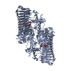
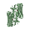
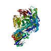
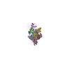
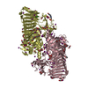
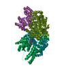
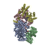
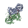
| Title | Structure of tetrameric HIV-1 Strand Transfer Complex Intasome | ||||||||||||
 Components Components |
| ||||||||||||
 Keywords Keywords |  VIRAL PROTEIN / integrase / integration / transposase / transesterification VIRAL PROTEIN / integrase / integration / transposase / transesterification | ||||||||||||
| Function / homology |  Function and homology information Function and homology informationRNA endonuclease activity / nucleotidyltransferase activity / HIV-1 retropepsin / : / retroviral ribonuclease H / exoribonuclease H / : / exoribonuclease H activity / host multivesicular body / DNA integration ...RNA endonuclease activity / nucleotidyltransferase activity / HIV-1 retropepsin / : / retroviral ribonuclease H / exoribonuclease H / : / exoribonuclease H activity / host multivesicular body / DNA integration / RNA-directed DNA polymerase / viral genome integration into host DNA / viral penetration into host nucleus / establishment of integrated proviral latency / RNA-directed DNA polymerase activity / Transferases; Transferring phosphorus-containing groups; Nucleotidyltransferases / RNA-DNA hybrid ribonuclease activity / viral nucleocapsid / endonuclease activity / DNA recombination / Hydrolases; Acting on ester bonds / nucleic acid binding / DNA-directed DNA polymerase / aspartic-type endopeptidase activity / DNA-directed DNA polymerase activity / symbiont entry into host cell / symbiont-mediated suppression of host gene expression / lipid binding / host cell nucleus / structural molecule activity / host cell plasma membrane / virion membrane / proteolysis / DNA binding / RNA binding / zinc ion binding / membraneSimilarity search - Function | ||||||||||||
| Biological species |   Sulfolobus solfataricus (archaea) Sulfolobus solfataricus (archaea)  Human immunodeficiency virus 1 Human immunodeficiency virus 1 Homo sapiens (human) Homo sapiens (human) | ||||||||||||
| Method | ELECTRON MICROSCOPY / single particle reconstruction / cryo EM / Resolution: 3.9 Å | ||||||||||||
 Authors Authors | Lyumkis, D. / Passos, D. | ||||||||||||
| Funding support |  United States, 3items United States, 3items
| ||||||||||||
 Citation Citation | Journal: Science / Year: 2017 Title: Cryo-EM structures and atomic model of the HIV-1 strand transfer complex intasome. Authors: Dario Oliveira Passos / Min Li / Renbin Yang / Stephanie V Rebensburg / Rodolfo Ghirlando / Youngmin Jeon / Nikoloz Shkriabai / Mamuka Kvaratskhelia / Robert Craigie / Dmitry Lyumkis / Abstract: Like all retroviruses, HIV-1 irreversibly inserts a viral DNA (vDNA) copy of its RNA genome into host target DNA (tDNA). The intasome, a higher-order nucleoprotein complex composed of viral integrase ...Like all retroviruses, HIV-1 irreversibly inserts a viral DNA (vDNA) copy of its RNA genome into host target DNA (tDNA). The intasome, a higher-order nucleoprotein complex composed of viral integrase (IN) and the ends of linear vDNA, mediates integration. Productive integration into host chromatin results in the formation of the strand transfer complex (STC) containing catalytically joined vDNA and tDNA. HIV-1 intasomes have been refractory to high-resolution structural studies. We used a soluble IN fusion protein to facilitate structural studies, through which we present a high-resolution cryo-electron microscopy (cryo-EM) structure of the core tetrameric HIV-1 STC and a higher-order form that adopts carboxyl-terminal domain rearrangements. The distinct STC structures highlight how HIV-1 can use the common retroviral intasome core architecture to accommodate different IN domain modules for assembly. | ||||||||||||
| History |
|
- Structure visualization
| Movie |
Movie viewer |
|---|---|
| Structure viewer | Molecule: MolmilJmol/JSmol |
- Downloads & links
-Download
| PDBx/mmCIF format | 5u1c.cif.gz | 212.4 KB | Display | PDBx/mmCIF format |
|---|---|---|---|---|
| PDB format | pdb5u1c.ent.gz | 156.5 KB | Display | PDB format |
| PDBx/mmJSON format | 5u1c.json.gz | Tree view | PDBx/mmJSON format | |
| Others |  Other downloads Other downloads |
-Validation report
| Arichive directory | https://data.pdbj.org/pub/pdb/validation_reports/u1/5u1cftp://data.pdbj.org/pub/pdb/validation_reports/u1/5u1c | HTTPS FTP |
|---|
-Related structure data
| Related structure data | 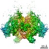 8481MC 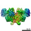 8483C M: map data used to model this data C: citing same article ( |
|---|---|
| Similar structure data |
-Links
 PDBj
PDBj


- Assembly
| Deposited unit | 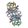
|
|---|---|
| 1 |
|
-Components
-Protein , 1 types, 4 molecules ABCD
| #1: Protein | Mass: 42320.273 Da / Num. of mol.: 4 / Mutation: E152Q Source method: isolated from a genetically manipulated source Source: (gene. exp.) Sulfolobus solfataricus (archaea), (gene. exp.) Human immunodeficiency virus 1Gene: SSOP1_2677, pol / Production host:  Escherichia coli (E. coli) Escherichia coli (E. coli)References: UniProt: A0A157T5S7, UniProt: F2WR39, UniProt: P12497*PLUS |
|---|
-DNA chain , 3 types, 6 molecules GHEIFJ
| #2: DNA chain | Mass: 3349.197 Da / Num. of mol.: 2 / Source method: obtained synthetically / Source: (synth.) Homo sapiens (human)#3: DNA chain | Mass: 7015.546 Da / Num. of mol.: 2 / Source method: obtained synthetically / Source: (synth.) Human immunodeficiency virus 1#4: DNA chain | Mass: 11414.358 Da / Num. of mol.: 2 / Source method: obtained synthetically Source: (synth.) Human immunodeficiency virus 1, (synth.) Homo sapiens (human) |
|---|
-Non-polymers , 2 types, 4 molecules 

| #5: Chemical |  Mass: 65.409 Da / Num. of mol.: 2 / Source method: obtained synthetically / Formula: Zn Mass: 65.409 Da / Num. of mol.: 2 / Source method: obtained synthetically / Formula: Zn#6: Chemical |  Mass: 24.305 Da / Num. of mol.: 2 / Source method: obtained synthetically / Formula: Mg Mass: 24.305 Da / Num. of mol.: 2 / Source method: obtained synthetically / Formula: Mg |
|---|
-Details
| Sequence details | The protein is a chimera of Sso7d linked to the N-terminus of integrase via an 11-glycine linker. |
|---|
-Experimental details
-Experiment
| Experiment | Method: ELECTRON MICROSCOPY |
|---|---|
| EM experiment | Aggregation state: PARTICLE / 3D reconstruction method: single particle reconstruction |
- Sample preparation
| Component | Name: complex formed by a tetrameric assembly of Sso7d-fusion HIV-1 Integrase with the product of DNA strand transfer Type: COMPLEX / Entity ID: #1-#4 / Source: RECOMBINANT | |||||||||||||||||||||||||
|---|---|---|---|---|---|---|---|---|---|---|---|---|---|---|---|---|---|---|---|---|---|---|---|---|---|---|
| Molecular weight | Value: 0.228 MDa / Experimental value: YES | |||||||||||||||||||||||||
| Source (natural) | Organism: Human immunodeficiency virus 1 | |||||||||||||||||||||||||
| Source (recombinant) | Organism: Escherichia coli (E. coli) / Plasmid: pSca355 | |||||||||||||||||||||||||
| Buffer solution | pH: 8 | |||||||||||||||||||||||||
| Buffer component |
| |||||||||||||||||||||||||
| Specimen | Conc.: 0.5 mg/ml / Embedding applied: NO / Shadowing applied: NO / Staining applied: NO / Vitrification applied: YES | |||||||||||||||||||||||||
| Specimen support | Grid material: GOLD / Grid mesh size: 400 divisions/in. / Grid type: Quantifoil | |||||||||||||||||||||||||
| Vitrification | Instrument: HOMEMADE PLUNGER / Cryogen name: ETHANE / Humidity: 50 % / Chamber temperature: 277 K Details: Sample containing HIV STC intasomes in SEC buffer was applied onto freshly plasma-treated (6 seconds, Gatan Solarus plasma cleaner) holey gold UltrAuFoil grids (Quantifoil), adsorbed for 30 ...Details: Sample containing HIV STC intasomes in SEC buffer was applied onto freshly plasma-treated (6 seconds, Gatan Solarus plasma cleaner) holey gold UltrAuFoil grids (Quantifoil), adsorbed for 30 seconds, then plunged into liquid ethane using a manual cryo-plunger in an ambient environment of 4 degrees C. |
- Electron microscopy imaging
| Experimental equipment |  Model: Titan Krios / Image courtesy: FEI Company |
|---|---|
| Microscopy | Model: FEI TITAN KRIOS |
| Electron gun | Electron source: FIELD EMISSION GUN / Accelerating voltage: 300 kV / Illumination mode: FLOOD BEAM |
| Electron lens | Mode: BRIGHT FIELDBright-field microscopy / Nominal magnification: 22500 X / Calibrated magnification: 38167 X / Calibrated defocus min: 1500 nm / Calibrated defocus max: 4000 nm / Cs: 2.7 mm / C2 aperture diameter: 100 µm / Alignment procedure: COMA FREE |
| Specimen holder | Cryogen: NITROGEN / Specimen holder model: FEI TITAN KRIOS AUTOGRID HOLDER / Temperature (max): 90 K / Temperature (min): 90 K |
| Image recording | Average exposure time: 20 sec. / Electron dose: 95 e/Å2 / Detector mode: COUNTING / Film or detector model: GATAN K2 SUMMIT (4k x 4k) / Num. of grids imaged: 1 / Num. of real images: 1225 Details: Individual frames were gain-corrected, aligned, and summed with the application of an exposure filter using MotionCor2, according to the nominal dose rate. |
| Image scans | Sampling size: 5 µm / Width: 3838 / Height: 3710 / Movie frames/image: 100 / Used frames/image: 1-100 |
- Processing
| Software | Name: PHENIX / Version: dev_2499: / Classification: refinement | ||||||||||||||||||||||||||||||||||||||||||||||||||||||||||||
|---|---|---|---|---|---|---|---|---|---|---|---|---|---|---|---|---|---|---|---|---|---|---|---|---|---|---|---|---|---|---|---|---|---|---|---|---|---|---|---|---|---|---|---|---|---|---|---|---|---|---|---|---|---|---|---|---|---|---|---|---|---|
| EM software |
| ||||||||||||||||||||||||||||||||||||||||||||||||||||||||||||
| CTF correction | Details: performed internally in Relion and Frealign / Type: PHASE FLIPPING AND AMPLITUDE CORRECTION | ||||||||||||||||||||||||||||||||||||||||||||||||||||||||||||
| Particle selection | Num. of particles selected: 274764 | ||||||||||||||||||||||||||||||||||||||||||||||||||||||||||||
| Symmetry | Point symmetry: C2 (2 fold cyclic) | ||||||||||||||||||||||||||||||||||||||||||||||||||||||||||||
| 3D reconstruction | Resolution: 3.9 Å / Resolution method: FSC 0.143 CUT-OFF / Num. of particles: 83766 / Algorithm: FOURIER SPACE / Details: Resolution-limited refinement used throughout / Symmetry type: POINT | ||||||||||||||||||||||||||||||||||||||||||||||||||||||||||||
| Atomic model building | B value: 180 / Protocol: FLEXIBLE FIT / Space: REAL / Target criteria: FSC 0.5 Details: To generate ensemble models, the complete intasome model was iteratively relaxed - using two-fold symmetry and a combination of Rosetta and Phenix - against one half map (the working map) ...Details: To generate ensemble models, the complete intasome model was iteratively relaxed - using two-fold symmetry and a combination of Rosetta and Phenix - against one half map (the working map) and inspected for consistency with the second half map (the free map). The model was then adjusted manually using Coot. Final ensemble modeling used half maps for all aspects of refinement and evaluation: 500 models were generated as described using Rosetta. From the 100 top-scoring models (scored by Rosetta energy), the ten models with the best map-to-model FSC were selected and refined in real space using secondary-structure restraints in Phenix. Molprobity was used throughout the refinement process. |
