 Movie
Movie Controller

[English] 日本語
 Yorodumi
- PDB-5u09: High-resolution crystal structure of the human CB1 cannabinoid re... -
+ Open data

- Basic information
| Entry | Database: PDB / ID: 5u09 | |||||||||
|---|---|---|---|---|---|---|---|---|---|---|
| Title | High-resolution crystal structure of the human CB1 cannabinoid receptor | |||||||||
 Components Components | Cannabinoid receptor 1,GlgA glycogen synthase | |||||||||
 Keywords Keywords |  MEMBRANE PROTEIN / helix MEMBRANE PROTEIN / helix | |||||||||
| Function / homology |  Function and homology information Function and homology informationcannabinoid signaling pathway / regulation of penile erection / retrograde trans-synaptic signaling by endocannabinoid / cannabinoid receptor activity / negative regulation of mast cell activation / trans-synaptic signaling by endocannabinoid, modulating synaptic transmission / glycogen (starch) synthase activity / negative regulation of fatty acid beta-oxidation / negative regulation of dopamine secretion / positive regulation of acute inflammatory response to antigenic stimulus ...cannabinoid signaling pathway / regulation of penile erection / retrograde trans-synaptic signaling by endocannabinoid / cannabinoid receptor activity / negative regulation of mast cell activation / trans-synaptic signaling by endocannabinoid, modulating synaptic transmission / glycogen (starch) synthase activity / negative regulation of fatty acid beta-oxidation / negative regulation of dopamine secretion / positive regulation of acute inflammatory response to antigenic stimulus / regulation of feeding behavior / negative regulation of serotonin secretion / regulation of presynaptic cytosolic calcium ion concentration / negative regulation of action potential / Class A/1 (Rhodopsin-like receptors) / positive regulation of blood pressure / regulation of metabolic process / positive regulation of fever generation / axonal fasciculation / regulation of synaptic transmission, GABAergic / regulation of insulin secretion / G protein-coupled receptor signaling pathway, coupled to cyclic nucleotide second messenger / GABA-ergic synapse / maternal process involved in female pregnancy / regulation of synaptic transmission, glutamatergic / negative regulation of blood pressure / response to nutrient / response to cocaine / G protein-coupled receptor activity / response to nicotine / adenylate cyclase-modulating G protein-coupled receptor signaling pathway / adenylate cyclase-activating G protein-coupled receptor signaling pathway / memory / positive regulation of neuron projection development / actin cytoskeleton / glucose homeostasis / presynaptic membrane / G alpha (i) signalling events / growth cone / spermatogenesis / response to ethanol / mitochondrial outer membrane / response to lipopolysaccharide / membrane raft / positive regulation of apoptotic process / nucleotide binding / glutamatergic synapse / identical protein binding / plasma membrane / cytoplasmSimilarity search - Function | |||||||||
| Biological species |  Homo sapiens (human) Homo sapiens (human)  Pyrococcus abyssi (archaea) Pyrococcus abyssi (archaea) | |||||||||
| Method | X-RAY DIFFRACTION / SYNCHROTRON / MOLECULAR REPLACEMENT / Resolution: 2.6 Å | |||||||||
 Authors Authors | Shao, Z.H. / Yin, J. / Rosenbaum, D. | |||||||||
 Citation Citation | Journal: Nature / Year: 2016 Title: High-resolution crystal structure of the human CB1 cannabinoid receptor. Authors: Shao, Z. / Yin, J. / Chapman, K. / Grzemska, M. / Clark, L. / Wang, J. / Rosenbaum, D.M. | |||||||||
| History |
|
- Structure visualization
| Structure viewer | Molecule: MolmilJmol/JSmol |
|---|
- Downloads & links
-Download
| PDBx/mmCIF format | 5u09.cif.gz | 116.2 KB | Display | PDBx/mmCIF format |
|---|---|---|---|---|
| PDB format | pdb5u09.ent.gz | 85.9 KB | Display | PDB format |
| PDBx/mmJSON format | 5u09.json.gz | Tree view | PDBx/mmJSON format | |
| Others |  Other downloads Other downloads |
-Validation report
| Arichive directory | https://data.pdbj.org/pub/pdb/validation_reports/u0/5u09ftp://data.pdbj.org/pub/pdb/validation_reports/u0/5u09 | HTTPS FTP |
|---|
-Related structure data
| Related structure data |  3v2yS S: Starting model for refinement |
|---|---|
| Similar structure data |


-Links
 PDBj
PDBj


- Assembly
| Deposited unit | 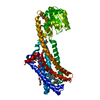
| ||||||||
|---|---|---|---|---|---|---|---|---|---|
| 1 |
| ||||||||
| Unit cell |
|
-Components
| #1: Protein | Mass: 57395.387 Da / Num. of mol.: 1 Fragment: P21554 residues 90-301, 333-421 and Q9V2J8 residues 218-413 Mutation: T210A Source method: isolated from a genetically manipulated source Source: (gene. exp.) Homo sapiens (human), (gene. exp.) Pyrococcus abyssi (strain GE5 / Orsay) (archaea)Gene: CNR1, CNR, PAB2292 / Plasmid: PFASTbac / Strain: GE5 / Orsay / Production host:   Spodoptera frugiperda (fall armyworm) / Strain (production host): sf9 / References: UniProt: P21554, UniProt: Q9V2J8 Spodoptera frugiperda (fall armyworm) / Strain (production host): sf9 / References: UniProt: P21554, UniProt: Q9V2J8 | ||||||
|---|---|---|---|---|---|---|---|
| #2: Chemical | ChemComp-PEG / Diethylene glycol  Mass: 106.120 Da / Num. of mol.: 9 / Source method: obtained synthetically / Formula: C4H10O3 Mass: 106.120 Da / Num. of mol.: 9 / Source method: obtained synthetically / Formula: C4H10O3#3: Chemical | ChemComp-SO4 / Sulfate  Mass: 96.063 Da / Num. of mol.: 4 / Source method: obtained synthetically / Formula: SO4 Mass: 96.063 Da / Num. of mol.: 4 / Source method: obtained synthetically / Formula: SO4#4: Chemical | ChemComp-7DY / | Taranabant  Mass: 515.955 Da / Num. of mol.: 1 / Source method: obtained synthetically / Formula: C27H25ClF3N3O2 / Comment: agonist*YM Mass: 515.955 Da / Num. of mol.: 1 / Source method: obtained synthetically / Formula: C27H25ClF3N3O2 / Comment: agonist*YM#5: Water | ChemComp-HOH / | Water Mass: 18.015 Da / Num. of mol.: 62 / Source method: isolated from a natural source / Formula: H2O Mass: 18.015 Da / Num. of mol.: 62 / Source method: isolated from a natural source / Formula: H2O |
-Experimental details
-Experiment
| Experiment | Method: X-RAY DIFFRACTION / Number of used crystals: 1 |
|---|
- Sample preparation
| Crystal | Density Matthews: 3.06 Å3/Da / Density % sol: 59.83 % |
|---|---|
| Crystal grow | Temperature: 293 K / Method: lipidic cubic phase / pH: 5.5 Details: 31% PEG400, 100mM Sodium Citrate pH5.5, 100mM magnesium sulfate |
-Data collection
| Diffraction | Mean temperature: 100 K | ||||||||||||||||||||||||||||||||||||||||||||||||||||||||||||||||||
|---|---|---|---|---|---|---|---|---|---|---|---|---|---|---|---|---|---|---|---|---|---|---|---|---|---|---|---|---|---|---|---|---|---|---|---|---|---|---|---|---|---|---|---|---|---|---|---|---|---|---|---|---|---|---|---|---|---|---|---|---|---|---|---|---|---|---|---|
| Diffraction source | Source: SYNCHROTRON / Site: APS  / Beamline: 23-ID-D / Wavelength: 1.033 Å / Beamline: 23-ID-D / Wavelength: 1.033 Å | ||||||||||||||||||||||||||||||||||||||||||||||||||||||||||||||||||
| Detector | Type: DECTRIS PILATUS 6M / Detector: PIXEL / Date: Jul 31, 2016 | ||||||||||||||||||||||||||||||||||||||||||||||||||||||||||||||||||
| Radiation | Protocol: SINGLE WAVELENGTH / Monochromatic (M) / Laue (L): M / Scattering type: x-ray | ||||||||||||||||||||||||||||||||||||||||||||||||||||||||||||||||||
| Radiation wavelength | Wavelength: 1.033 Å / Relative weight: 1 | ||||||||||||||||||||||||||||||||||||||||||||||||||||||||||||||||||
| Reflection | Resolution: 2.6→50.01 Å / Num. obs: 19781 / % possible obs: 96.9 % / Redundancy: 5.4 % / Rmerge(I) obs: 0.194 / Net I/av σ(I): 7.432 / Net I/σ(I): 5.6 | ||||||||||||||||||||||||||||||||||||||||||||||||||||||||||||||||||
| Reflection shell |
|
- Processing
| Software |
| |||||||||||||||||||||||||||||||||||||||||||||||||||||||||||||||||||||||||||
|---|---|---|---|---|---|---|---|---|---|---|---|---|---|---|---|---|---|---|---|---|---|---|---|---|---|---|---|---|---|---|---|---|---|---|---|---|---|---|---|---|---|---|---|---|---|---|---|---|---|---|---|---|---|---|---|---|---|---|---|---|---|---|---|---|---|---|---|---|---|---|---|---|---|---|---|---|
| Refinement | Method to determine structure: MOLECULAR REPLACEMENT Starting model: 3V2Y Resolution: 2.6→50.01 Å / Cor.coef. Fo:Fc: 0.928 / Cor.coef. Fo:Fc free: 0.88 / SU B: 10.364 / SU ML: 0.22 / Cross valid method: THROUGHOUT / σ(F): 0 / ESU R Free: 0.452 Details: HYDROGENS HAVE BEEN ADDED IN THE RIDING POSITIONS U VALUES : REFINED INDIVIDUALLY. AS PER THE AUTHORS THE NUMBER OF OBSERVED REFLECTIONS USED FOR REFINEMENT IS LOW DUE TO ANISOTROPIC TRUNCATION OF THE DATA.
| |||||||||||||||||||||||||||||||||||||||||||||||||||||||||||||||||||||||||||
| Solvent computation | Ion probe radii: 0.8 Å / Shrinkage radii: 0.8 Å / VDW probe radii: 1.2 Å | |||||||||||||||||||||||||||||||||||||||||||||||||||||||||||||||||||||||||||
| Displacement parameters | Biso max: 151.65 Å2 / Biso mean: 44.12 Å2 / Biso min: 7.02 Å2
| |||||||||||||||||||||||||||||||||||||||||||||||||||||||||||||||||||||||||||
| Refinement step | Cycle: final / Resolution: 2.6→50.01 Å
| |||||||||||||||||||||||||||||||||||||||||||||||||||||||||||||||||||||||||||
| Refine LS restraints |
| |||||||||||||||||||||||||||||||||||||||||||||||||||||||||||||||||||||||||||
| LS refinement shell | Resolution: 2.598→2.665 Å / Total num. of bins used: 20
|
