 Movie
Movie Controller


+ Open data

- Basic information
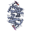
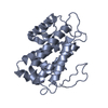
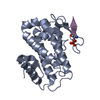
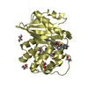
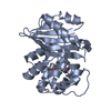
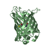
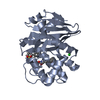
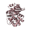
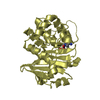
| Entry | Database: PDB / ID: 5twg | ||||||
|---|---|---|---|---|---|---|---|
| Title | human MOB1A bound to human MST1 phosphorylated T353 peptide | ||||||
 Components Components |
| ||||||
 Keywords Keywords |  SIGNALING PROTEIN / complex SIGNALING PROTEIN / complex | ||||||
| Function / homology |  Function and homology information Function and homology informationpositive regulation of hepatocyte apoptotic process / cell differentiation involved in embryonic placenta development / regulation of cell differentiation involved in embryonic placenta development / primitive hemopoiesis / neural tube formation / positive regulation of extrinsic apoptotic signaling pathway via death domain receptors / positive regulation of substrate-dependent cell migration, cell attachment to substrate / endocardium development / negative regulation of organ growth / hippo signaling ...positive regulation of hepatocyte apoptotic process / cell differentiation involved in embryonic placenta development / regulation of cell differentiation involved in embryonic placenta development / primitive hemopoiesis / neural tube formation / positive regulation of extrinsic apoptotic signaling pathway via death domain receptors / positive regulation of substrate-dependent cell migration, cell attachment to substrate / endocardium development / negative regulation of organ growth / hippo signaling / Signaling by Hippo / organ growth / branching involved in blood vessel morphogenesis / protein kinase activator activity / hepatocyte apoptotic process / regulation of MAPK cascade / extrinsic apoptotic signaling pathway via death domain receptors / positive regulation of fat cell differentiation / canonical Wnt signaling pathway / keratinocyte differentiation / protein serine/threonine kinase activator activity / epithelial cell proliferation / central nervous system development / protein tetramerization / negative regulation of canonical Wnt signaling pathway / cell morphogenesis / protein import into nucleus / negative regulation of epithelial cell proliferation / positive regulation of protein binding / positive regulation of peptidyl-serine phosphorylation / peptidyl-serine phosphorylation / RNA polymerase II-specific DNA-binding transcription factor binding / protein autophosphorylation / nuclear body / protein stabilization / non-specific serine/threonine protein kinase / protein kinase activity / intracellular signal transduction / positive regulation of apoptotic process / positive regulation of protein phosphorylation / protein phosphorylation / protein serine kinase activity / protein serine/threonine kinase activity / apoptotic process / magnesium ion binding / signal transduction / protein homodimerization activity / protein-containing complex / extracellular exosome / nucleoplasm / ATP binding / identical protein binding / metal ion binding / nucleus / cytosol / cytoplasmSimilarity search - Function | ||||||
| Biological species |  Homo sapiens (human) Homo sapiens (human) | ||||||
| Method | X-RAY DIFFRACTION / SYNCHROTRON / Resolution: 2.3 Å | ||||||
 Authors Authors | Xiong, S. / Sicheri, F. | ||||||
 Citation Citation | Journal: Mol. Cell Proteomics / Year: 2017 Title: MOB1 Mediated Phospho-recognition in the Core Mammalian Hippo Pathway. Authors: Couzens, A.L. / Xiong, S. / Knight, J.D.R. / Mao, D.Y. / Guettler, S. / Picaud, S. / Kurinov, I. / Filippakopoulos, P. / Sicheri, F. / Gingras, A.C. | ||||||
| History |
|
- Structure visualization
| Structure viewer | Molecule: MolmilJmol/JSmol |
|---|
- Downloads & links
-Download
| PDBx/mmCIF format | 5twg.cif.gz | 92.3 KB | Display | PDBx/mmCIF format |
|---|---|---|---|---|
| PDB format | pdb5twg.ent.gz | 74.9 KB | Display | PDB format |
| PDBx/mmJSON format | 5twg.json.gz | Tree view | PDBx/mmJSON format | |
| Others |  Other downloads Other downloads |
-Validation report
| Arichive directory | https://data.pdbj.org/pub/pdb/validation_reports/tw/5twgftp://data.pdbj.org/pub/pdb/validation_reports/tw/5twg | HTTPS FTP |
|---|
-Related structure data


-Links
 PDBj
PDBj


- Assembly
| Deposited unit | 
| ||||||||||||
|---|---|---|---|---|---|---|---|---|---|---|---|---|---|
| 1 |
| ||||||||||||
| Unit cell |
| ||||||||||||
| Components on special symmetry positions |
|
-Components
| #1: Protein | Mass: 25111.754 Da / Num. of mol.: 1 Source method: isolated from a genetically manipulated source Source: (gene. exp.) Homo sapiens (human) / Gene: MOB1A, C2orf6, MOB4B, MOBK1B, MOBKL1B / Production host:  Escherichia coli (E. coli) / References: UniProt: Q9H8S9 Escherichia coli (E. coli) / References: UniProt: Q9H8S9 |
|---|---|
| #2: Protein/peptide | Mass: 1617.691 Da / Num. of mol.: 1 Source method: isolated from a genetically manipulated source Source: (gene. exp.) Homo sapiens (human) / Production host: Escherichia coli (E. coli) / References: UniProt: Q13043*PLUS |
| #3: Chemical | ChemComp-ZN /   Mass: 65.409 Da / Num. of mol.: 1 / Source method: obtained synthetically / Formula: Zn Mass: 65.409 Da / Num. of mol.: 1 / Source method: obtained synthetically / Formula: Zn |
| #4: Water | ChemComp-HOH / Water Mass: 18.015 Da / Num. of mol.: 23 / Source method: isolated from a natural source / Formula: H2O Mass: 18.015 Da / Num. of mol.: 23 / Source method: isolated from a natural source / Formula: H2O |
-Experimental details
-Experiment
| Experiment | Method: X-RAY DIFFRACTION / Number of used crystals: 1 |
|---|
- Sample preparation
| Crystal | Density Matthews: 2.48 Å3/Da / Density % sol: 50.33 % |
|---|---|
| Crystal grow | Temperature: 298 K / Method: vapor diffusion / Details: 0.1 M MES pH 6.0, 0.2 M LiCl and 20% PEG 6000 |
-Data collection
| Diffraction | Mean temperature: 100 K |
|---|---|
| Diffraction source | Source: SYNCHROTRON / Site: APS  / Beamline: 24-ID-C / Wavelength: 0.9792 Å / Beamline: 24-ID-C / Wavelength: 0.9792 Å |
| Detector | Type: DECTRIS PILATUS 6M-F / Detector: PIXEL / Date: Feb 5, 2015 |
| Radiation | Protocol: SINGLE WAVELENGTH / Monochromatic (M) / Laue (L): M / Scattering type: x-ray |
| Radiation wavelength | Wavelength: 0.9792 Å / Relative weight: 1 |
| Reflection | Resolution: 2.1→56 Å / Num. obs: 11982 / % possible obs: 96.5 % / Redundancy: 2.8 % / Net I/σ(I): 30 |
- Processing
| Software |
| ||||||||||||||||||||||||||||||||||||||||||||||||||||||||||||||||||||||||||||||||||||||||||||||||||||||||||||||||||||||||||||||||||||||||||||||||||||||||||||||||||||||||||||||||||||||||||||||||||||||||||||||||||||||||||||||||||||||||||||||||||||||||||
|---|---|---|---|---|---|---|---|---|---|---|---|---|---|---|---|---|---|---|---|---|---|---|---|---|---|---|---|---|---|---|---|---|---|---|---|---|---|---|---|---|---|---|---|---|---|---|---|---|---|---|---|---|---|---|---|---|---|---|---|---|---|---|---|---|---|---|---|---|---|---|---|---|---|---|---|---|---|---|---|---|---|---|---|---|---|---|---|---|---|---|---|---|---|---|---|---|---|---|---|---|---|---|---|---|---|---|---|---|---|---|---|---|---|---|---|---|---|---|---|---|---|---|---|---|---|---|---|---|---|---|---|---|---|---|---|---|---|---|---|---|---|---|---|---|---|---|---|---|---|---|---|---|---|---|---|---|---|---|---|---|---|---|---|---|---|---|---|---|---|---|---|---|---|---|---|---|---|---|---|---|---|---|---|---|---|---|---|---|---|---|---|---|---|---|---|---|---|---|---|---|---|---|---|---|---|---|---|---|---|---|---|---|---|---|---|---|---|---|---|---|---|---|---|---|---|---|---|---|---|---|---|---|---|---|---|---|---|---|---|---|---|---|---|---|---|---|---|---|---|---|---|
| Refinement | Resolution: 2.3→45.602 Å / SU ML: 0.33 / Cross valid method: FREE R-VALUE / σ(F): 1.34 / Phase error: 29.4 / Stereochemistry target values: ML
| ||||||||||||||||||||||||||||||||||||||||||||||||||||||||||||||||||||||||||||||||||||||||||||||||||||||||||||||||||||||||||||||||||||||||||||||||||||||||||||||||||||||||||||||||||||||||||||||||||||||||||||||||||||||||||||||||||||||||||||||||||||||||||
| Solvent computation | Shrinkage radii: 0.9 Å / VDW probe radii: 1.11 Å / Solvent model: FLAT BULK SOLVENT MODEL | ||||||||||||||||||||||||||||||||||||||||||||||||||||||||||||||||||||||||||||||||||||||||||||||||||||||||||||||||||||||||||||||||||||||||||||||||||||||||||||||||||||||||||||||||||||||||||||||||||||||||||||||||||||||||||||||||||||||||||||||||||||||||||
| Refinement step | Cycle: LAST / Resolution: 2.3→45.602 Å
| ||||||||||||||||||||||||||||||||||||||||||||||||||||||||||||||||||||||||||||||||||||||||||||||||||||||||||||||||||||||||||||||||||||||||||||||||||||||||||||||||||||||||||||||||||||||||||||||||||||||||||||||||||||||||||||||||||||||||||||||||||||||||||
| Refine LS restraints |
| ||||||||||||||||||||||||||||||||||||||||||||||||||||||||||||||||||||||||||||||||||||||||||||||||||||||||||||||||||||||||||||||||||||||||||||||||||||||||||||||||||||||||||||||||||||||||||||||||||||||||||||||||||||||||||||||||||||||||||||||||||||||||||
| LS refinement shell |
| ||||||||||||||||||||||||||||||||||||||||||||||||||||||||||||||||||||||||||||||||||||||||||||||||||||||||||||||||||||||||||||||||||||||||||||||||||||||||||||||||||||||||||||||||||||||||||||||||||||||||||||||||||||||||||||||||||||||||||||||||||||||||||
| Refinement TLS params. | Method: refined / Refine-ID: X-RAY DIFFRACTION
| ||||||||||||||||||||||||||||||||||||||||||||||||||||||||||||||||||||||||||||||||||||||||||||||||||||||||||||||||||||||||||||||||||||||||||||||||||||||||||||||||||||||||||||||||||||||||||||||||||||||||||||||||||||||||||||||||||||||||||||||||||||||||||
| Refinement TLS group |
|
