 Movie
Movie Controller


+ Open data

- Basic information
| Entry | Database: PDB / ID: 5tnu | ||||||
|---|---|---|---|---|---|---|---|
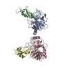
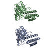
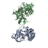
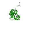
| Title | S. tokodaii XPB II crystal structure at 3.0 Angstrom resolution | ||||||
 Components Components | DNA-dependent ATPase XPBII | ||||||
 Keywords Keywords |  TRANSCRIPTION / Helicase / NER TRANSCRIPTION / Helicase / NER | ||||||
| Function / homology |  Function and homology information Function and homology information | ||||||
| Biological species |   Sulfolobus tokodaii (archaea) Sulfolobus tokodaii (archaea) | ||||||
| Method | X-RAY DIFFRACTION / SYNCHROTRON / MOLECULAR REPLACEMENT / molecular replacement / Resolution: 3.05 Å | ||||||
 Authors Authors | DuPrez, K.T. / Hilario, E. / Wang, I. / Fan, L. | ||||||
| Funding support |  United States, 1items United States, 1items
| ||||||
 Citation Citation | Journal: Anal.Chem. / Year: 2018 Title: Application of Electrochemical Devices to Characterize the Dynamic Actions of Helicases on DNA. Authors: Kahanda, D. / DuPrez, K.T. / Hilario, E. / McWilliams, M.A. / Wohlgamuth, C.H. / Fan, L. / Slinker, J.D. | ||||||
| History |
|
- Structure visualization
| Structure viewer | Molecule: MolmilJmol/JSmol |
|---|
- Downloads & links
-Download
| PDBx/mmCIF format | 5tnu.cif.gz | 185.3 KB | Display | PDBx/mmCIF format |
|---|---|---|---|---|
| PDB format | pdb5tnu.ent.gz | 142.9 KB | Display | PDB format |
| PDBx/mmJSON format | 5tnu.json.gz | Tree view | PDBx/mmJSON format | |
| Others |  Other downloads Other downloads |
-Validation report
| Arichive directory | https://data.pdbj.org/pub/pdb/validation_reports/tn/5tnuftp://data.pdbj.org/pub/pdb/validation_reports/tn/5tnu | HTTPS FTP |
|---|
-Related structure data
| Related structure data | 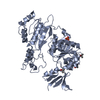 2fwrS S: Starting model for refinement |
|---|---|
| Similar structure data |
-Links
 PDBj
PDBj- Assembly
| Deposited unit | 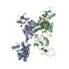
| ||||||||||||||||||
|---|---|---|---|---|---|---|---|---|---|---|---|---|---|---|---|---|---|---|---|
| 1 | 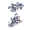
| ||||||||||||||||||
| 2 | 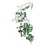
| ||||||||||||||||||
| Unit cell |
| ||||||||||||||||||
| Noncrystallographic symmetry (NCS) | NCS domain:
NCS domain segments: Component-ID: 0 / Ens-ID: 1 / Beg auth comp-ID: SER / Beg label comp-ID: SER / End auth comp-ID: GLU / End label comp-ID: GLU / Refine code: 0 / Auth seq-ID: -1 - 422 / Label seq-ID: 19 - 442
|
-Components
| #1: Protein | Mass: 52887.230 Da / Num. of mol.: 2 Source method: isolated from a genetically manipulated source Source: (gene. exp.) Sulfolobus tokodaii (strain DSM 16993 / JCM 10545 / NBRC 100140 / 7) (archaea)Strain: DSM 16993 / JCM 10545 / NBRC 100140 / 7 / Gene: xpb2, ST1613, STK_16130 / Plasmid: pET-15b / Production host:  Escherichia coli BL21(DE3) (bacteria) / Strain (production host): BL21 / Variant (production host): DE3 pLysS Rosetta / References: UniProt: Q970I2 Escherichia coli BL21(DE3) (bacteria) / Strain (production host): BL21 / Variant (production host): DE3 pLysS Rosetta / References: UniProt: Q970I2#2: Chemical | ChemComp-SO4 / Sulfate  Mass: 96.063 Da / Num. of mol.: 12 / Source method: obtained synthetically / Formula: SO4 Mass: 96.063 Da / Num. of mol.: 12 / Source method: obtained synthetically / Formula: SO4#3: Chemical | ChemComp-CL / Chloride  Mass: 35.453 Da / Num. of mol.: 7 / Source method: obtained synthetically / Formula: Cl Mass: 35.453 Da / Num. of mol.: 7 / Source method: obtained synthetically / Formula: Cl#4: Chemical | ChemComp-GOL / Glycerol  Mass: 92.094 Da / Num. of mol.: 7 / Source method: obtained synthetically / Formula: C3H8O3 Mass: 92.094 Da / Num. of mol.: 7 / Source method: obtained synthetically / Formula: C3H8O3#5: Water | ChemComp-HOH / | Water Mass: 18.015 Da / Num. of mol.: 129 / Source method: isolated from a natural source / Formula: H2O Mass: 18.015 Da / Num. of mol.: 129 / Source method: isolated from a natural source / Formula: H2O |
|---|
-Experimental details
-Experiment
| Experiment | Method: X-RAY DIFFRACTION / Number of used crystals: 1 |
|---|
- Sample preparation
| Crystal | Density Matthews: 3.9 Å3/Da / Density % sol: 68.48 % |
|---|---|
| Crystal grow | Temperature: 298 K / Method: vapor diffusion, hanging drop / pH: 5.6 Details: 1:1 ratio of protein with 10 mM sodium citrate pH 5.6, 1770 mM ammonium sulfate |
-Data collection
| Diffraction | Mean temperature: 100 K | ||||||||||||||||||||||||||||||
|---|---|---|---|---|---|---|---|---|---|---|---|---|---|---|---|---|---|---|---|---|---|---|---|---|---|---|---|---|---|---|---|
| Diffraction source | Source: SYNCHROTRON / Site: ALS / Beamline: 12.3.1 / Wavelength: 1 Å | ||||||||||||||||||||||||||||||
| Detector | Type: ADSC QUANTUM 315 / Detector: CCD / Date: Feb 22, 2015 | ||||||||||||||||||||||||||||||
| Radiation | Protocol: SINGLE WAVELENGTH / Monochromatic (M) / Laue (L): M / Scattering type: x-ray | ||||||||||||||||||||||||||||||
| Radiation wavelength | Wavelength: 1 Å / Relative weight: 1 | ||||||||||||||||||||||||||||||
| Reflection | Resolution: 3.05→29.88 Å / Num. obs: 31341 / % possible obs: 99.9 % / Redundancy: 23.6 % / CC1/2: 0.996 / Rmerge(I) obs: 0.374 / Rpim(I) all: 0.078 / Rrim(I) all: 0.382 / Net I/σ(I): 11.2 / Num. measured all: 740378 / Scaling rejects: 440 | ||||||||||||||||||||||||||||||
| Reflection shell | Diffraction-ID: 1
|
-Phasing
| Phasing | Method: molecular replacement | ||||||||||||||||||||||||||||||||||||||||||||||||||||||||||||||||||||||||||||||||||||||||||||||||||||||||||||||||||||
|---|---|---|---|---|---|---|---|---|---|---|---|---|---|---|---|---|---|---|---|---|---|---|---|---|---|---|---|---|---|---|---|---|---|---|---|---|---|---|---|---|---|---|---|---|---|---|---|---|---|---|---|---|---|---|---|---|---|---|---|---|---|---|---|---|---|---|---|---|---|---|---|---|---|---|---|---|---|---|---|---|---|---|---|---|---|---|---|---|---|---|---|---|---|---|---|---|---|---|---|---|---|---|---|---|---|---|---|---|---|---|---|---|---|---|---|---|---|
| Phasing MR | Model details: Phaser MODE: MR_AUTO
| ||||||||||||||||||||||||||||||||||||||||||||||||||||||||||||||||||||||||||||||||||||||||||||||||||||||||||||||||||||
| Phasing dm | Method: Solvent flattening and Histogram matching / Reflection: 31287 | ||||||||||||||||||||||||||||||||||||||||||||||||||||||||||||||||||||||||||||||||||||||||||||||||||||||||||||||||||||
| Phasing dm shell |
|
- Processing
| Software |
| ||||||||||||||||||||||||||||||||||||||||||||||||||||||||||||
|---|---|---|---|---|---|---|---|---|---|---|---|---|---|---|---|---|---|---|---|---|---|---|---|---|---|---|---|---|---|---|---|---|---|---|---|---|---|---|---|---|---|---|---|---|---|---|---|---|---|---|---|---|---|---|---|---|---|---|---|---|---|
| Refinement | Method to determine structure: MOLECULAR REPLACEMENT Starting model: 2FWR Resolution: 3.05→29.88 Å / Cor.coef. Fo:Fc: 0.939 / Cor.coef. Fo:Fc free: 0.914 / WRfactor Rfree: 0.1998 / WRfactor Rwork: 0.1745 / FOM work R set: 0.7979 / SU B: 17.044 / SU ML: 0.292 / SU R Cruickshank DPI: 0.8848 / SU Rfree: 0.3317 / Cross valid method: THROUGHOUT / σ(F): 0 / ESU R: 0.885 / ESU R Free: 0.332 / Stereochemistry target values: MAXIMUM LIKELIHOOD Details: HYDROGENS HAVE BEEN ADDED IN THE RIDING POSITIONS U VALUES : REFINED INDIVIDUALLY
| ||||||||||||||||||||||||||||||||||||||||||||||||||||||||||||
| Solvent computation | Ion probe radii: 0.8 Å / Shrinkage radii: 0.8 Å / VDW probe radii: 1.2 Å / Solvent model: MASK | ||||||||||||||||||||||||||||||||||||||||||||||||||||||||||||
| Displacement parameters | Biso max: 328.86 Å2 / Biso mean: 90.199 Å2 / Biso min: 19.48 Å2
| ||||||||||||||||||||||||||||||||||||||||||||||||||||||||||||
| Refinement step | Cycle: final / Resolution: 3.05→29.88 Å
| ||||||||||||||||||||||||||||||||||||||||||||||||||||||||||||
| Refine LS restraints |
| ||||||||||||||||||||||||||||||||||||||||||||||||||||||||||||
| Refine LS restraints NCS | Ens-ID: 1 / Number: 20024 / Refine-ID: X-RAY DIFFRACTION / Type: interatomic distance / Rms dev position: 0.19 Å / Weight position: 0.05
| ||||||||||||||||||||||||||||||||||||||||||||||||||||||||||||
| LS refinement shell | Resolution: 3.05→3.128 Å / Rfactor Rfree error: 0 / Total num. of bins used: 20
|
