 Movie
Movie Controller


+ Open data

- Basic information
| Entry | Database: PDB / ID: 5tmh | ||||||
|---|---|---|---|---|---|---|---|
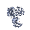
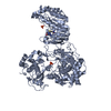
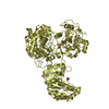
| Title | Structure of Zika virus NS5 | ||||||
 Components Components | Polyprotein Proteolysis Proteolysis | ||||||
 Keywords Keywords | VIRAL PROTEIN | ||||||
| Function / homology |  Function and homology information Function and homology informationsymbiont-mediated suppression of host JAK-STAT cascade via inhibition of host TYK2 activity / flavivirin / symbiont-mediated suppression of host JAK-STAT cascade via inhibition of STAT2 activity / symbiont-mediated suppression of host JAK-STAT cascade via inhibition of STAT1 activity / ribonucleoside triphosphate phosphatase activity / double-stranded RNA binding / viral capsid / nucleoside-triphosphate phosphatase / symbiont-mediated suppression of host cytoplasmic pattern recognition receptor signaling pathway via inhibition of TBK1 activity / symbiont-mediated suppression of host toll-like receptor signaling pathway ...symbiont-mediated suppression of host JAK-STAT cascade via inhibition of host TYK2 activity / flavivirin / symbiont-mediated suppression of host JAK-STAT cascade via inhibition of STAT2 activity / symbiont-mediated suppression of host JAK-STAT cascade via inhibition of STAT1 activity / ribonucleoside triphosphate phosphatase activity / double-stranded RNA binding / viral capsid / nucleoside-triphosphate phosphatase / symbiont-mediated suppression of host cytoplasmic pattern recognition receptor signaling pathway via inhibition of TBK1 activity / symbiont-mediated suppression of host toll-like receptor signaling pathway / mRNA (guanine-N7)-methyltransferase / methyltransferase cap1 / clathrin-dependent endocytosis of virus by host cell / mRNA (nucleoside-2'-O-)-methyltransferase activity / mRNA 5'-cap (guanine-N7-)-methyltransferase activity / RNA helicase activity / host cell perinuclear region of cytoplasm / host cell endoplasmic reticulum membrane / protein dimerization activity / symbiont-mediated suppression of host type I interferon-mediated signaling pathway / RNA helicase / symbiont entry into host cell / induction by virus of host autophagy / RNA-directed RNA polymerase / virus-mediated perturbation of host defense response / viral RNA genome replication / RNA-dependent RNA polymerase activity / serine-type endopeptidase activity / fusion of virus membrane with host endosome membrane / viral envelope / lipid binding / host cell nucleus / virion attachment to host cell / GTP binding / virion membrane / structural molecule activity / ATP hydrolysis activity / proteolysis / extracellular region / ATP binding / membrane / metal ion bindingSimilarity search - Function | ||||||
| Biological species |   Zika virus Zika virus | ||||||
| Method | X-RAY DIFFRACTION / SYNCHROTRON / Resolution: 3.278 Å | ||||||
 Authors Authors | Wang, B. / Tan, X. / Song, J. | ||||||
 Citation Citation | Journal: Nat Commun / Year: 2017 Title: The structure of Zika virus NS5 reveals a conserved domain conformation. Authors: Wang, B. / Tan, X.F. / Thurmond, S. / Zhang, Z.M. / Lin, A. / Hai, R. / Song, J. | ||||||
| History |
|
- Structure visualization
| Structure viewer | Molecule: MolmilJmol/JSmol |
|---|
- Downloads & links
-Download
| PDBx/mmCIF format | 5tmh.cif.gz | 672.1 KB | Display | PDBx/mmCIF format |
|---|---|---|---|---|
| PDB format | pdb5tmh.ent.gz | 552.1 KB | Display | PDB format |
| PDBx/mmJSON format | 5tmh.json.gz | Tree view | PDBx/mmJSON format | |
| Others |  Other downloads Other downloads |
-Validation report
| Arichive directory | https://data.pdbj.org/pub/pdb/validation_reports/tm/5tmhftp://data.pdbj.org/pub/pdb/validation_reports/tm/5tmh | HTTPS FTP |
|---|
-Related structure data
| Similar structure data |
|---|


-Links
 PDBj
PDBj


- Assembly
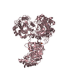
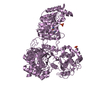
| Deposited unit | 
| ||||||||
|---|---|---|---|---|---|---|---|---|---|
| 1 | 
| ||||||||
| 2 | 
| ||||||||
| Unit cell |
|
-Components
| #1: Protein | Proteolysis Mass: 103005.547 Da / Num. of mol.: 2 Source method: isolated from a genetically manipulated source Source: (gene. exp.) Zika virus (strain Mr 766) / Strain: Mr 766Production host: References: UniProt: A0A192GPS6, UniProt: Q32ZE1*PLUS #2: Chemical | S-Adenosyl-L-homocysteine  Type: L-peptide linking / Mass: 384.411 Da / Num. of mol.: 2 Type: L-peptide linking / Mass: 384.411 Da / Num. of mol.: 2Source method: isolated from a genetically manipulated source Formula: C14H20N6O5S #3: Chemical | ChemComp-ZN /   Mass: 65.409 Da / Num. of mol.: 4 Mass: 65.409 Da / Num. of mol.: 4Source method: isolated from a genetically manipulated source Formula: Zn #4: Chemical | Sulfate  Mass: 96.063 Da / Num. of mol.: 2 Mass: 96.063 Da / Num. of mol.: 2Source method: isolated from a genetically manipulated source Formula: SO4 #5: Chemical | ChemComp-GOL / Glycerol  Mass: 92.094 Da / Num. of mol.: 4 Mass: 92.094 Da / Num. of mol.: 4Source method: isolated from a genetically manipulated source Formula: C3H8O3 |
|---|
-Experimental details
-Experiment
| Experiment | Method: X-RAY DIFFRACTION / Number of used crystals: 1 |
|---|
- Sample preparation
| Crystal | Density Matthews: 3.12 Å3/Da / Density % sol: 60.64 % |
|---|---|
| Crystal grow | Temperature: 277.15 K / Method: vapor diffusion, hanging drop / Details: 0.7-0.9 M lithium sulfate, 0.1 M MES, pH 6-7 |
-Data collection
| Diffraction | Mean temperature: 277 K |
|---|---|
| Diffraction source | Source: SYNCHROTRON / Site: ALS  / Beamline: 5.0.3 / Wavelength: 0.9774 Å / Beamline: 5.0.3 / Wavelength: 0.9774 Å |
| Detector | Type: ADSC QUANTUM 315r / Detector: CCD / Date: Sep 24, 2016 |
| Radiation | Protocol: SINGLE WAVELENGTH / Monochromatic (M) / Laue (L): M / Scattering type: x-ray |
| Radiation wavelength | Wavelength: 0.9774 Å / Relative weight: 1 |
| Reflection | Resolution: 3.278→50 Å / Num. obs: 39593 / % possible obs: 99.4 % / Redundancy: 6.2 % / Net I/σ(I): 5.3 |
- Processing
| Software |
| ||||||||||||||||||||||||||||||||||||||||||||||||||||||||||||||||||||||||||||||||||||||||||||||||||||||||||||||||||||||||||||||||||||||||||||||||||||||||||||||||||||||||||||||||||||||||||||||||||||||||
|---|---|---|---|---|---|---|---|---|---|---|---|---|---|---|---|---|---|---|---|---|---|---|---|---|---|---|---|---|---|---|---|---|---|---|---|---|---|---|---|---|---|---|---|---|---|---|---|---|---|---|---|---|---|---|---|---|---|---|---|---|---|---|---|---|---|---|---|---|---|---|---|---|---|---|---|---|---|---|---|---|---|---|---|---|---|---|---|---|---|---|---|---|---|---|---|---|---|---|---|---|---|---|---|---|---|---|---|---|---|---|---|---|---|---|---|---|---|---|---|---|---|---|---|---|---|---|---|---|---|---|---|---|---|---|---|---|---|---|---|---|---|---|---|---|---|---|---|---|---|---|---|---|---|---|---|---|---|---|---|---|---|---|---|---|---|---|---|---|---|---|---|---|---|---|---|---|---|---|---|---|---|---|---|---|---|---|---|---|---|---|---|---|---|---|---|---|---|---|---|---|---|
| Refinement | Resolution: 3.278→49.244 Å / SU ML: 0.42 / Cross valid method: FREE R-VALUE / σ(F): 1.33 / Phase error: 34.76 / Stereochemistry target values: ML
| ||||||||||||||||||||||||||||||||||||||||||||||||||||||||||||||||||||||||||||||||||||||||||||||||||||||||||||||||||||||||||||||||||||||||||||||||||||||||||||||||||||||||||||||||||||||||||||||||||||||||
| Solvent computation | Shrinkage radii: 0.9 Å / VDW probe radii: 1.11 Å / Solvent model: FLAT BULK SOLVENT MODEL | ||||||||||||||||||||||||||||||||||||||||||||||||||||||||||||||||||||||||||||||||||||||||||||||||||||||||||||||||||||||||||||||||||||||||||||||||||||||||||||||||||||||||||||||||||||||||||||||||||||||||
| Refinement step | Cycle: LAST / Resolution: 3.278→49.244 Å
| ||||||||||||||||||||||||||||||||||||||||||||||||||||||||||||||||||||||||||||||||||||||||||||||||||||||||||||||||||||||||||||||||||||||||||||||||||||||||||||||||||||||||||||||||||||||||||||||||||||||||
| Refine LS restraints |
| ||||||||||||||||||||||||||||||||||||||||||||||||||||||||||||||||||||||||||||||||||||||||||||||||||||||||||||||||||||||||||||||||||||||||||||||||||||||||||||||||||||||||||||||||||||||||||||||||||||||||
| LS refinement shell |
| ||||||||||||||||||||||||||||||||||||||||||||||||||||||||||||||||||||||||||||||||||||||||||||||||||||||||||||||||||||||||||||||||||||||||||||||||||||||||||||||||||||||||||||||||||||||||||||||||||||||||
| Refinement TLS params. | Method: refined / Refine-ID: X-RAY DIFFRACTION
| ||||||||||||||||||||||||||||||||||||||||||||||||||||||||||||||||||||||||||||||||||||||||||||||||||||||||||||||||||||||||||||||||||||||||||||||||||||||||||||||||||||||||||||||||||||||||||||||||||||||||
| Refinement TLS group |
|
