 Movie
Movie Controller


+ Open data

- Basic information
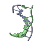
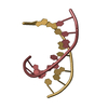
| Entry | Database: PDB / ID: 5tko | ||||||
|---|---|---|---|---|---|---|---|
| Title | RNA heptamer duplex with one 2'-5'-linkage | ||||||
 Components Components |
| ||||||
 Keywords Keywords |  RNA / 2'-5'-LINKAGE RNA / 2'-5'-LINKAGE | ||||||
| Function / homology | : / RNA Function and homology information Function and homology information | ||||||
| Biological species | unidentified (others) | ||||||
| Method | X-RAY DIFFRACTION / SYNCHROTRON / MOLECULAR REPLACEMENT / Resolution: 1.52 Å | ||||||
 Authors Authors | Luo, Z. / Sheng, J. | ||||||
 Citation Citation | Journal: To Be Published Title: RNA heptamer duplex with one 2'-5'-linkage Authors: Luo, Z. / Sheng, J. / Wang, R. | ||||||
| History |
|
- Structure visualization
| Structure viewer | Molecule: MolmilJmol/JSmol |
|---|
- Downloads & links
-Download
| PDBx/mmCIF format | 5tko.cif.gz | 45.5 KB | Display | PDBx/mmCIF format |
|---|---|---|---|---|
| PDB format | pdb5tko.ent.gz | 32.1 KB | Display | PDB format |
| PDBx/mmJSON format | 5tko.json.gz | Tree view | PDBx/mmJSON format | |
| Others |  Other downloads Other downloads |
-Validation report
| Arichive directory | https://data.pdbj.org/pub/pdb/validation_reports/tk/5tkoftp://data.pdbj.org/pub/pdb/validation_reports/tk/5tko | HTTPS FTP |
|---|
-Related structure data
| Related structure data |  4u37S S: Starting model for refinement |
|---|---|
| Similar structure data |


-Links
 PDBj
PDBj
- Assembly
| Deposited unit | 
| ||||||||
|---|---|---|---|---|---|---|---|---|---|
| 1 | 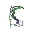
| ||||||||
| 2 | 
| ||||||||
| Unit cell |
|
-Components
| #1: RNA chain | Mass: 2157.331 Da / Num. of mol.: 2 / Source method: obtained synthetically / Source: (synth.) unidentified (others) #2: RNA chain | Mass: 2260.419 Da / Num. of mol.: 2 / Source method: obtained synthetically / Source: (synth.) unidentified (others) #3: Chemical | ChemComp-CO / |   Mass: 58.933 Da / Num. of mol.: 1 / Source method: obtained synthetically / Formula: Co Mass: 58.933 Da / Num. of mol.: 1 / Source method: obtained synthetically / Formula: Co#4: Water | ChemComp-HOH / | Water Mass: 18.015 Da / Num. of mol.: 87 / Source method: isolated from a natural source / Formula: H2O Mass: 18.015 Da / Num. of mol.: 87 / Source method: isolated from a natural source / Formula: H2O |
|---|
-Experimental details
-Experiment
| Experiment | Method: X-RAY DIFFRACTION / Number of used crystals: 1 |
|---|
- Sample preparation
| Crystal | Density Matthews: 2.19 Å3/Da / Density % sol: 43.92 % |
|---|---|
| Crystal grow | Temperature: 293 K / Method: vapor diffusion, sitting drop / pH: 5.5 Details: Precipitant: 40 mM sodium cacodylate pH5.5, 10% (v/v) MPD, 20 mM hexamine cobalt (III) chloride, 20mM MgCl2 Reservoir: 35% (v/v) MPD |
-Data collection
| Diffraction | Mean temperature: 100 K |
|---|---|
| Diffraction source | Source: SYNCHROTRON / Site: APS  / Beamline: 22-ID / Wavelength: 1 Å / Beamline: 22-ID / Wavelength: 1 Å |
| Detector | Type: RAYONIX MX300-HS / Detector: CCD / Date: Dec 5, 2015 |
| Radiation | Protocol: SINGLE WAVELENGTH / Monochromatic (M) / Laue (L): M / Scattering type: x-ray |
| Radiation wavelength | Wavelength: 1 Å / Relative weight: 1 |
| Reflection | Resolution: 1.52→30 Å / Num. obs: 11869 / % possible obs: 99.8 % / Redundancy: 6.1 % / Rmerge(I) obs: 0.083 / Net I/σ(I): 17.8 |
| Reflection shell | Resolution: 1.52→1.57 Å / Redundancy: 5.7 % / Mean I/σ(I) obs: 1.95 / % possible all: 100 |
- Processing
| Software |
| ||||||||||||||||||||||||||||||||||||||||||||||||||||||||||||||||||||||||||||||||||||||||||||||||||||||||||||||||||||||||||||||||||||||||||||||||||||||||||||||||||||||||||||||||||||||
|---|---|---|---|---|---|---|---|---|---|---|---|---|---|---|---|---|---|---|---|---|---|---|---|---|---|---|---|---|---|---|---|---|---|---|---|---|---|---|---|---|---|---|---|---|---|---|---|---|---|---|---|---|---|---|---|---|---|---|---|---|---|---|---|---|---|---|---|---|---|---|---|---|---|---|---|---|---|---|---|---|---|---|---|---|---|---|---|---|---|---|---|---|---|---|---|---|---|---|---|---|---|---|---|---|---|---|---|---|---|---|---|---|---|---|---|---|---|---|---|---|---|---|---|---|---|---|---|---|---|---|---|---|---|---|---|---|---|---|---|---|---|---|---|---|---|---|---|---|---|---|---|---|---|---|---|---|---|---|---|---|---|---|---|---|---|---|---|---|---|---|---|---|---|---|---|---|---|---|---|---|---|---|---|
| Refinement | Method to determine structure: MOLECULAR REPLACEMENT Starting model: 4U37 Resolution: 1.52→27.58 Å / Cor.coef. Fo:Fc: 0.972 / Cor.coef. Fo:Fc free: 0.962 / SU B: 2.904 / SU ML: 0.046 / Cross valid method: THROUGHOUT / ESU R: 0.099 / ESU R Free: 0.075 / Details: HYDROGENS HAVE BEEN ADDED IN THE RIDING POSITIONS
| ||||||||||||||||||||||||||||||||||||||||||||||||||||||||||||||||||||||||||||||||||||||||||||||||||||||||||||||||||||||||||||||||||||||||||||||||||||||||||||||||||||||||||||||||||||||
| Solvent computation | Ion probe radii: 0.8 Å / Shrinkage radii: 0.8 Å / VDW probe radii: 1.2 Å | ||||||||||||||||||||||||||||||||||||||||||||||||||||||||||||||||||||||||||||||||||||||||||||||||||||||||||||||||||||||||||||||||||||||||||||||||||||||||||||||||||||||||||||||||||||||
| Displacement parameters | Biso mean: 16.635 Å2
| ||||||||||||||||||||||||||||||||||||||||||||||||||||||||||||||||||||||||||||||||||||||||||||||||||||||||||||||||||||||||||||||||||||||||||||||||||||||||||||||||||||||||||||||||||||||
| Refinement step | Cycle: 1 / Resolution: 1.52→27.58 Å
| ||||||||||||||||||||||||||||||||||||||||||||||||||||||||||||||||||||||||||||||||||||||||||||||||||||||||||||||||||||||||||||||||||||||||||||||||||||||||||||||||||||||||||||||||||||||
| Refine LS restraints |
|