 Movie
Movie Controller


+ Open data

- Basic information
| Entry | Database: PDB / ID: 5tiz | ||||||
|---|---|---|---|---|---|---|---|
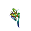
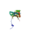
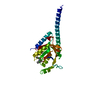
| Title | Schistosoma japonicum (Blood Fluke) Sulfotransferase | ||||||
 Components Components | Sulfotransferase | ||||||
 Keywords Keywords | TRANSFERASE / SULFOTRANSFERASE / PARASITE / HELMINTH | ||||||
| Function / homology | Sulfotransferase, S. mansonii-type / Sulfotransferase domain / P-loop containing nucleotide triphosphate hydrolases / Rossmann fold / P-loop containing nucleoside triphosphate hydrolase / 3-Layer(aba) Sandwich / Alpha Beta / ADENOSINE-3'-5'-DIPHOSPHATE / Uncharacterized protein Function and homology information Function and homology information | ||||||
| Biological species |  | ||||||
| Method |  X-RAY DIFFRACTION / SYNCHROTRON / MOLECULAR REPLACEMENT / Resolution: 2.87 Å X-RAY DIFFRACTION / SYNCHROTRON / MOLECULAR REPLACEMENT / Resolution: 2.87 Å | ||||||
 Authors Authors | Taylor, A.B. / Hart, P.J. | ||||||
 Citation Citation | Journal: J. Biol. Chem. / Year: 2017 Title: Structural and enzymatic insights into species-specific resistance to schistosome parasite drug therapy. Authors: Taylor, A.B. / Roberts, K.M. / Cao, X. / Clark, N.E. / Holloway, S.P. / Donati, E. / Polcaro, C.M. / Pica-Mattoccia, L. / Tarpley, R.S. / McHardy, S.F. / Cioli, D. / LoVerde, P.T. / ...Authors: Taylor, A.B. / Roberts, K.M. / Cao, X. / Clark, N.E. / Holloway, S.P. / Donati, E. / Polcaro, C.M. / Pica-Mattoccia, L. / Tarpley, R.S. / McHardy, S.F. / Cioli, D. / LoVerde, P.T. / Fitzpatrick, P.F. / Hart, P.J. | ||||||
| History |
|
- Structure visualization
| Structure viewer | Molecule: MolmilJmol/JSmol |
|---|
- Downloads & links
-Download
| PDBx/mmCIF format | 5tiz.cif.gz | 65.4 KB | Display | PDBx/mmCIF format |
|---|---|---|---|---|
| PDB format | pdb5tiz.ent.gz | 46.5 KB | Display | PDB format |
| PDBx/mmJSON format | 5tiz.json.gz | Tree view | PDBx/mmJSON format | |
| Others |  Other downloads Other downloads |
-Validation report
| Summary document | 5tiz_validation.pdf.gz | 778.1 KB | Display | wwPDB validaton report |
|---|---|---|---|---|
| Full document | 5tiz_full_validation.pdf.gz | 778.9 KB | Display | |
| Data in XML | 5tiz_validation.xml.gz | 10.9 KB | Display | |
| Data in CIF | 5tiz_validation.cif.gz | 13.6 KB | Display | |
| Arichive directory | https://data.pdbj.org/pub/pdb/validation_reports/ti/5tizftp://data.pdbj.org/pub/pdb/validation_reports/ti/5tiz | HTTPS FTP |
-Related structure data
| Related structure data |  5tivC  5tiwC  5tixC  5tiyC 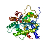 4muaS C: citing same article ( S: Starting model for refinement |
|---|---|
| Similar structure data |


-Links
 PDBj
PDBj
- Assembly
| Deposited unit | 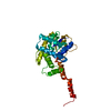
| ||||||||
|---|---|---|---|---|---|---|---|---|---|
| 1 |
| ||||||||
| Unit cell |
|
-Components
| #1: Protein | Mass: 30598.033 Da / Num. of mol.: 1 Source method: isolated from a genetically manipulated source Source: (gene. exp.)  |
|---|---|
| #2: Chemical | ChemComp-A3P /   Type: RNA linking / Mass: 427.201 Da / Num. of mol.: 1 / Source method: obtained synthetically / Formula: C10H15N5O10P2 Type: RNA linking / Mass: 427.201 Da / Num. of mol.: 1 / Source method: obtained synthetically / Formula: C10H15N5O10P2 |
-Experimental details
-Experiment
| Experiment | Method: X-RAY DIFFRACTION / Number of used crystals: 1 |
|---|
- Sample preparation
| Crystal | Density Matthews: 2.43 Å3/Da / Density % sol: 49.48 % |
|---|---|
| Crystal grow | Temperature: 295 K / Method: vapor diffusion, sitting drop Details: 20%(v/v) PEG 8000, 3%(v/v) MPD, 0.1M Imidazole/Hydrochloric acid pH 6.5 |
-Data collection
| Diffraction | Mean temperature: 100 K |
|---|---|
| Diffraction source | Source: SYNCHROTRON / Site: APS  / Beamline: 24-ID-E / Wavelength: 0.97918 Å / Beamline: 24-ID-E / Wavelength: 0.97918 Å |
| Detector | Type: ADSC QUANTUM 315 / Detector: CCD / Date: Nov 16, 2015 |
| Radiation | Protocol: SINGLE WAVELENGTH / Monochromatic (M) / Laue (L): M / Scattering type: x-ray |
| Radiation wavelength | Wavelength: 0.97918 Å / Relative weight: 1 |
| Reflection | Resolution: 2.87→54.65 Å / Num. obs: 7110 / % possible obs: 96.9 % / Redundancy: 2.8 % / Biso Wilson estimate: 39 Å2 / Rsym value: 0.158 / Net I/σ(I): 7.7 |
| Reflection shell | Resolution: 2.87→3.03 Å / Redundancy: 2.8 % / Rmerge(I) obs: 0.667 / Mean I/σ(I) obs: 2 / % possible all: 98.8 |
- Processing
| Software |
| ||||||||||||||||||||||||||||||||||||||||||
|---|---|---|---|---|---|---|---|---|---|---|---|---|---|---|---|---|---|---|---|---|---|---|---|---|---|---|---|---|---|---|---|---|---|---|---|---|---|---|---|---|---|---|---|
| Refinement | Method to determine structure: MOLECULAR REPLACEMENT Starting model: 4MUA Resolution: 2.87→54.648 Å / SU ML: 0.45 / Cross valid method: THROUGHOUT / σ(F): 1.36 / Phase error: 25.94
| ||||||||||||||||||||||||||||||||||||||||||
| Solvent computation | Shrinkage radii: 0.9 Å / VDW probe radii: 1.11 Å | ||||||||||||||||||||||||||||||||||||||||||
| Displacement parameters | Biso mean: 31.4 Å2 | ||||||||||||||||||||||||||||||||||||||||||
| Refinement step | Cycle: LAST / Resolution: 2.87→54.648 Å
| ||||||||||||||||||||||||||||||||||||||||||
| Refine LS restraints |
| ||||||||||||||||||||||||||||||||||||||||||
| LS refinement shell |
|
