 Movie
Movie Controller

[English] 日本語
 Yorodumi
- PDB-5thl: Crystal structure of the human tyrosyl-tRNA synthetase mutant G41R -
+ Open data

- Basic information
| Entry | Database: PDB / ID: 5thl | ||||||
|---|---|---|---|---|---|---|---|
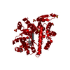
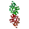
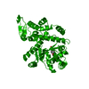
| Title | Crystal structure of the human tyrosyl-tRNA synthetase mutant G41R | ||||||
 Components Components | Tyrosine--tRNA ligase, cytoplasmic | ||||||
 Keywords Keywords |  LIGASE / Tyrosyl-tRNA synthetase / CMT / mutant LIGASE / Tyrosyl-tRNA synthetase / CMT / mutant | ||||||
| Function / homology |  Function and homology informationinterleukin-8 receptor binding / tyrosyl-tRNA aminoacylation / tyrosine-tRNA ligase / tyrosine-tRNA ligase activity / Cytosolic tRNA aminoacylation / response to starvation / small molecule binding / tRNA binding / nuclear body / apoptotic process ...interleukin-8 receptor binding / tyrosyl-tRNA aminoacylation / tyrosine-tRNA ligase / tyrosine-tRNA ligase activity / Cytosolic tRNA aminoacylation / response to starvation / small molecule binding / tRNA binding / nuclear body / apoptotic process / extracellular space / RNA binding / ATP binding / nucleus / cytosol / cytoplasm Function and homology informationinterleukin-8 receptor binding / tyrosyl-tRNA aminoacylation / tyrosine-tRNA ligase / tyrosine-tRNA ligase activity / Cytosolic tRNA aminoacylation / response to starvation / small molecule binding / tRNA binding / nuclear body / apoptotic process ...interleukin-8 receptor binding / tyrosyl-tRNA aminoacylation / tyrosine-tRNA ligase / tyrosine-tRNA ligase activity / Cytosolic tRNA aminoacylation / response to starvation / small molecule binding / tRNA binding / nuclear body / apoptotic process / extracellular space / RNA binding / ATP binding / nucleus / cytosol / cytoplasmSimilarity search - Function | ||||||
| Biological species |  Homo sapiens (human) Homo sapiens (human) | ||||||
| Method | X-RAY DIFFRACTION / SYNCHROTRON / MOLECULAR REPLACEMENT / Resolution: 1.6 Å | ||||||
 Authors Authors | Blocquel, D. / Sajish, M. / Yang, X.L. | ||||||
 Citation Citation | Journal: Nucleic Acids Res. / Year: 2017 Title: Alternative stable conformation capable of protein misinteraction links tRNA synthetase to peripheral neuropathy. Authors: Blocquel, D. / Li, S. / Wei, N. / Daub, H. / Sajish, M. / Erfurth, M.L. / Kooi, G. / Zhou, J. / Bai, G. / Schimmel, P. / Jordanova, A. / Yang, X.L. | ||||||
| History |
|
- Structure visualization
| Structure viewer | Molecule: MolmilJmol/JSmol |
|---|
- Downloads & links
-Download
| PDBx/mmCIF format | 5thl.cif.gz | 89 KB | Display | PDBx/mmCIF format |
|---|---|---|---|---|
| PDB format | pdb5thl.ent.gz | 64.7 KB | Display | PDB format |
| PDBx/mmJSON format | 5thl.json.gz | Tree view | PDBx/mmJSON format | |
| Others |  Other downloads Other downloads |
-Validation report
| Arichive directory | https://data.pdbj.org/pub/pdb/validation_reports/th/5thlftp://data.pdbj.org/pub/pdb/validation_reports/th/5thl | HTTPS FTP |
|---|
-Related structure data
| Related structure data |  5thhC 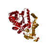 1n3lS C: citing same article ( S: Starting model for refinement |
|---|---|
| Similar structure data |

-Links
 PDBj
PDBj
- Assembly
| Deposited unit | 
| ||||||||
|---|---|---|---|---|---|---|---|---|---|
| 1 | 
| ||||||||
| Unit cell |
| ||||||||
| Components on special symmetry positions |
|
-Components
| #1: Protein | Mass: 42233.699 Da / Num. of mol.: 1 / Mutation: G41R Source method: isolated from a genetically manipulated source Source: (gene. exp.) Homo sapiens (human) / Gene: YARS / Production host:  Escherichia coli (E. coli) / References: UniProt: P54577, tyrosine-tRNA ligase Escherichia coli (E. coli) / References: UniProt: P54577, tyrosine-tRNA ligase |
|---|---|
| #2: Water | ChemComp-HOH / Water Mass: 18.015 Da / Num. of mol.: 387 / Source method: isolated from a natural source / Formula: H2O Mass: 18.015 Da / Num. of mol.: 387 / Source method: isolated from a natural source / Formula: H2O |
-Experimental details
-Experiment
| Experiment | Method: X-RAY DIFFRACTION / Number of used crystals: 1 |
|---|
- Sample preparation
| Crystal | Density Matthews: 2.53 Å3/Da / Density % sol: 51.35 % |
|---|---|
| Crystal grow | Temperature: 289 K / Method: vapor diffusion / Details: ammonium sulfate, acetone, sodium phosphate |
-Data collection
| Diffraction | Mean temperature: 193 K | |||||||||||||||||||||||||||||||||||||||||||||||||||||||||||||||||||||||||||||||||||||||||||||||||||||||||
|---|---|---|---|---|---|---|---|---|---|---|---|---|---|---|---|---|---|---|---|---|---|---|---|---|---|---|---|---|---|---|---|---|---|---|---|---|---|---|---|---|---|---|---|---|---|---|---|---|---|---|---|---|---|---|---|---|---|---|---|---|---|---|---|---|---|---|---|---|---|---|---|---|---|---|---|---|---|---|---|---|---|---|---|---|---|---|---|---|---|---|---|---|---|---|---|---|---|---|---|---|---|---|---|---|---|---|
| Diffraction source | Source: SYNCHROTRON / Site: SSRL  / Beamline: BL9-2 / Wavelength: 0.97945 Å / Beamline: BL9-2 / Wavelength: 0.97945 Å | |||||||||||||||||||||||||||||||||||||||||||||||||||||||||||||||||||||||||||||||||||||||||||||||||||||||||
| Detector | Type: DECTRIS PILATUS 6M / Detector: PIXEL / Date: Apr 1, 2011 | |||||||||||||||||||||||||||||||||||||||||||||||||||||||||||||||||||||||||||||||||||||||||||||||||||||||||
| Radiation | Protocol: SINGLE WAVELENGTH / Monochromatic (M) / Laue (L): M / Scattering type: x-ray | |||||||||||||||||||||||||||||||||||||||||||||||||||||||||||||||||||||||||||||||||||||||||||||||||||||||||
| Radiation wavelength | Wavelength: 0.97945 Å / Relative weight: 1 | |||||||||||||||||||||||||||||||||||||||||||||||||||||||||||||||||||||||||||||||||||||||||||||||||||||||||
| Reflection | Resolution: 1.6→50 Å / Num. obs: 57743 / % possible obs: 99.4 % / Redundancy: 6.8 % / Rmerge(I) obs: 0.077 / Χ2: 2.018 / Net I/av σ(I): 39.789 / Net I/σ(I): 11.4 / Num. measured all: 392235 | |||||||||||||||||||||||||||||||||||||||||||||||||||||||||||||||||||||||||||||||||||||||||||||||||||||||||
| Reflection shell |
|
- Processing
| Software |
| ||||||||||||||||||||||||
|---|---|---|---|---|---|---|---|---|---|---|---|---|---|---|---|---|---|---|---|---|---|---|---|---|---|
| Refinement | Method to determine structure: MOLECULAR REPLACEMENT Starting model: 1N3L Resolution: 1.6→31.989 Å / SU ML: 0.16 / Cross valid method: NONE / σ(F): 0 / Phase error: 19.18
| ||||||||||||||||||||||||
| Solvent computation | Shrinkage radii: 0.95 Å / VDW probe radii: 1.2 Å / Bsol: 37.304 Å2 / ksol: 0.382 e/Å3 | ||||||||||||||||||||||||
| Displacement parameters | Biso max: 71.87 Å2 / Biso mean: 24.9432 Å2 / Biso min: 10.33 Å2
| ||||||||||||||||||||||||
| Refinement step | Cycle: final / Resolution: 1.6→31.989 Å
|
