 Movie
Movie Controller

[English] 日本語
 Yorodumi
- PDB-5tg8: Crystal structure of H15 hemagglutinin from A/shearwater/WA/2576/... -
+ Open data

- Basic information
| Entry | Database: PDB / ID: 5tg8 | |||||||||
|---|---|---|---|---|---|---|---|---|---|---|
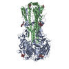
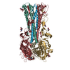
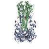
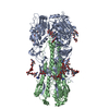
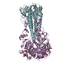
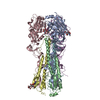
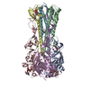
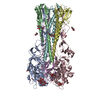
| Title | Crystal structure of H15 hemagglutinin from A/shearwater/WA/2576/1979 H15N9 influenza virus | |||||||||
 Components Components |
| |||||||||
 Keywords Keywords |  VIRAL PROTEIN / Influenza virus / hemagglutinin / HA / H15N9 / Receptor specificity VIRAL PROTEIN / Influenza virus / hemagglutinin / HA / H15N9 / Receptor specificity | |||||||||
| Function / homology |  Function and homology informationviral budding from plasma membrane / clathrin-dependent endocytosis of virus by host cell / host cell surface receptor binding / apical plasma membrane / fusion of virus membrane with host plasma membrane / fusion of virus membrane with host endosome membrane / viral envelope / virion attachment to host cell / host cell plasma membrane / virion membrane Function and homology informationviral budding from plasma membrane / clathrin-dependent endocytosis of virus by host cell / host cell surface receptor binding / apical plasma membrane / fusion of virus membrane with host plasma membrane / fusion of virus membrane with host endosome membrane / viral envelope / virion attachment to host cell / host cell plasma membrane / virion membraneSimilarity search - Function | |||||||||
| Biological species |   Influenza A virus Influenza A virus | |||||||||
| Method | X-RAY DIFFRACTION / SYNCHROTRON / MOLECULAR REPLACEMENT / Resolution: 3.1 Å | |||||||||
 Authors Authors | Wilson, I.A. / Tzarum, N. | |||||||||
| Funding support |  United States, 2items United States, 2items
| |||||||||
 Citation Citation | Journal: J. Virol. / Year: 2017 Title: Unique Structural Features of Influenza Virus H15 Hemagglutinin. Authors: Tzarum, N. / McBride, R. / Nycholat, C.M. / Peng, W. / Paulson, J.C. / Wilson, I.A. | |||||||||
| History |
|
- Structure visualization
| Structure viewer | Molecule: MolmilJmol/JSmol |
|---|
- Downloads & links
-Download
| PDBx/mmCIF format | 5tg8.cif.gz | 206.7 KB | Display | PDBx/mmCIF format |
|---|---|---|---|---|
| PDB format | pdb5tg8.ent.gz | 163.7 KB | Display | PDB format |
| PDBx/mmJSON format | 5tg8.json.gz | Tree view | PDBx/mmJSON format | |
| Others |  Other downloads Other downloads |
-Validation report
| Arichive directory | https://data.pdbj.org/pub/pdb/validation_reports/tg/5tg8ftp://data.pdbj.org/pub/pdb/validation_reports/tg/5tg8 | HTTPS FTP |
|---|
-Related structure data
| Related structure data |  5tg9C 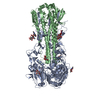 4n5jS C: citing same article ( S: Starting model for refinement |
|---|---|
| Similar structure data |


-Links
 PDBj
PDBj


- Assembly
| Deposited unit | 
| ||||||||
|---|---|---|---|---|---|---|---|---|---|
| 1 | 
| ||||||||
| 2 | 
| ||||||||
| Unit cell |
|
-Components
| #1: Protein | Mass: 36210.758 Da / Num. of mol.: 2 Source method: isolated from a genetically manipulated source Source: (gene. exp.) Influenza A virus (A/shearWater/Australia/2576/1979(H15N9))Strain: A/shearWater/Australia/2576/1979(H15N9) / Gene: HA / Production host:  Trichoplusia ni (cabbage looper) / References: UniProt: L0L3X3 Trichoplusia ni (cabbage looper) / References: UniProt: L0L3X3#2: Protein | Mass: 22210.434 Da / Num. of mol.: 2 Source method: isolated from a genetically manipulated source Source: (gene. exp.) Influenza A virus / Strain: A/shearWater/Australia/2576/1979(H15N9) / Gene: HA / Production host: Trichoplusia ni (cabbage looper) / References: UniProt: L0L3X3#3: Polysaccharide | / Mass: 424.401 Da / Num. of mol.: 2Source method: isolated from a genetically manipulated source #4: Sugar | N-Acetylglucosamine  Type: D-saccharide, beta linking / Mass: 221.208 Da / Num. of mol.: 2 Type: D-saccharide, beta linking / Mass: 221.208 Da / Num. of mol.: 2Source method: isolated from a genetically manipulated source Formula: C8H15NO6 |
|---|
-Experimental details
-Experiment
| Experiment | Method: X-RAY DIFFRACTION / Number of used crystals: 1 |
|---|
- Sample preparation
| Crystal | Density Matthews: 4.18 Å3/Da / Density % sol: 70.54 % |
|---|---|
| Crystal grow | Temperature: 293.15 K / Method: vapor diffusion, sitting drop / pH: 8 / Details: 20% (w/v) PEG 3500, 0.2M potassium nitrate pH 6.9. |
-Data collection
| Diffraction | Mean temperature: 100 K |
|---|---|
| Diffraction source | Source: SYNCHROTRON / Site: APS / Beamline: 23-ID-B / Wavelength: 1.0332 Å |
| Detector | Type: DECTRIS PILATUS 6M / Detector: PIXEL / Date: Jun 6, 2015 |
| Radiation | Protocol: SINGLE WAVELENGTH / Monochromatic (M) / Laue (L): M / Scattering type: x-ray |
| Radiation wavelength | Wavelength: 1.0332 Å / Relative weight: 1 |
| Reflection | Resolution: 3.1→50 Å / Num. obs: 34747 / % possible obs: 98.7 % / Redundancy: 4.1 % / Rsym value: 0.18 / Net I/σ(I): 9.1 |
| Reflection shell | Resolution: 3.1→3.15 Å / % possible obs: 90.7 % / Redundancy: 3.1 % / CC1/2: 0.894 / Net I/σ(I) obs: 1.7 |
- Processing
| Software |
| ||||||||||||||||||||||||||||||||||||||||||||||||||||||||||||||||||||||||||||||||||||||||||||||||||
|---|---|---|---|---|---|---|---|---|---|---|---|---|---|---|---|---|---|---|---|---|---|---|---|---|---|---|---|---|---|---|---|---|---|---|---|---|---|---|---|---|---|---|---|---|---|---|---|---|---|---|---|---|---|---|---|---|---|---|---|---|---|---|---|---|---|---|---|---|---|---|---|---|---|---|---|---|---|---|---|---|---|---|---|---|---|---|---|---|---|---|---|---|---|---|---|---|---|---|---|
| Refinement | Method to determine structure: MOLECULAR REPLACEMENT Starting model: 4N5J Resolution: 3.1→39.388 Å / SU ML: 0.44 / Cross valid method: FREE R-VALUE / σ(F): 1.96 / Phase error: 29.63
| ||||||||||||||||||||||||||||||||||||||||||||||||||||||||||||||||||||||||||||||||||||||||||||||||||
| Solvent computation | Shrinkage radii: 0.9 Å / VDW probe radii: 1.11 Å | ||||||||||||||||||||||||||||||||||||||||||||||||||||||||||||||||||||||||||||||||||||||||||||||||||
| Refinement step | Cycle: LAST / Resolution: 3.1→39.388 Å
| ||||||||||||||||||||||||||||||||||||||||||||||||||||||||||||||||||||||||||||||||||||||||||||||||||
| Refine LS restraints |
| ||||||||||||||||||||||||||||||||||||||||||||||||||||||||||||||||||||||||||||||||||||||||||||||||||
| LS refinement shell |
|
