 Movie
Movie Controller

[English] 日本語
 Yorodumi
- PDB-5tc6: Crystal structure of human 5'-deoxy-5'-methylthioadenosine phosph... -
+ Open data

- Basic information
| Entry | Database: PDB / ID: 5tc6 | ||||||
|---|---|---|---|---|---|---|---|
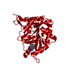
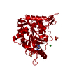
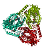
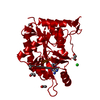
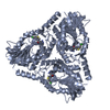
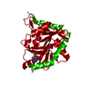
| Title | Crystal structure of human 5'-deoxy-5'-methylthioadenosine phosphorylase in complex with propylthio-immucillin-A | ||||||
 Components Components | S-methyl-5'-thioadenosine phosphorylase | ||||||
 Keywords Keywords | TRANSFERASE/TRANSFERASE INHIBITOR / phosphorylase / inhibitor / complex / TRANSFERASE-TRANSFERASE INHIBITOR complex | ||||||
| Function / homology |  Function and homology information Function and homology informationMethionine salvage pathway / S-methyl-5'-thioadenosine phosphorylase / 1,4-alpha-oligoglucan phosphorylase activity / S-methyl-5-thioadenosine phosphorylase activity / L-methionine salvage from methylthioadenosine / nucleobase-containing compound metabolic process / purine ribonucleoside salvage / response to testosterone / Gene and protein expression by JAK-STAT signaling after Interleukin-12 stimulation / methylation ...Methionine salvage pathway / S-methyl-5'-thioadenosine phosphorylase / 1,4-alpha-oligoglucan phosphorylase activity / S-methyl-5-thioadenosine phosphorylase activity / L-methionine salvage from methylthioadenosine / nucleobase-containing compound metabolic process / purine ribonucleoside salvage / response to testosterone / Gene and protein expression by JAK-STAT signaling after Interleukin-12 stimulation / methylation / extracellular exosome / nucleoplasm / cytosolSimilarity search - Function | ||||||
| Biological species |  Homo sapiens (human) Homo sapiens (human) | ||||||
| Method | X-RAY DIFFRACTION / SYNCHROTRON / MOLECULAR REPLACEMENT / molecular replacement / Resolution: 1.48 Å | ||||||
 Authors Authors | Cameron, S.A. / Firestone, R.S. / Schramm, V.L. / Almo, S.C. | ||||||
| Funding support |  United States, 1items United States, 1items
| ||||||
 Citation Citation | Journal: To be published Title: TBA Authors: Firestone, R.S. / Cameron, S.A. / Karp, J.M. / Arcus, V.L. / Schramm, V.L. | ||||||
| History |
|
- Structure visualization
| Structure viewer | Molecule: MolmilJmol/JSmol |
|---|
- Downloads & links
-Download
| PDBx/mmCIF format | 5tc6.cif.gz | 78.1 KB | Display | PDBx/mmCIF format |
|---|---|---|---|---|
| PDB format | pdb5tc6.ent.gz | 54.7 KB | Display | PDB format |
| PDBx/mmJSON format | 5tc6.json.gz | Tree view | PDBx/mmJSON format | |
| Others |  Other downloads Other downloads |
-Validation report
| Arichive directory | https://data.pdbj.org/pub/pdb/validation_reports/tc/5tc6ftp://data.pdbj.org/pub/pdb/validation_reports/tc/5tc6 | HTTPS FTP |
|---|
-Related structure data
| Related structure data | 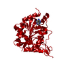 1k27S S: Starting model for refinement |
|---|---|
| Similar structure data |


-Links
 PDBj
PDBj


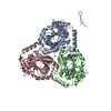
- Assembly
| Deposited unit | 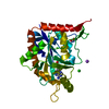
| ||||||||||||||||||
|---|---|---|---|---|---|---|---|---|---|---|---|---|---|---|---|---|---|---|---|
| 1 | 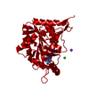
| ||||||||||||||||||
| Unit cell |
| ||||||||||||||||||
| Components on special symmetry positions |
|
-Components
-Protein , 1 types, 1 molecules A
| #1: Protein | / 5'-methylthioadenosine phosphorylase / MTAPase Mass: 33119.051 Da / Num. of mol.: 1 Source method: isolated from a genetically manipulated source Source: (gene. exp.) Homo sapiens (human) / Gene: MTAP, MSAP / Plasmid: pJexpress414 / Production host:  Escherichia coli (E. coli) / Strain (production host): BL21(DE3) Escherichia coli (E. coli) / Strain (production host): BL21(DE3)References: UniProt: Q13126, S-methyl-5'-thioadenosine phosphorylase |
|---|
-Non-polymers , 6 types, 227 molecules 


| #2: Chemical | ChemComp-7A6 / ( Mass: 323.414 Da / Num. of mol.: 1 / Source method: obtained synthetically / Formula: C14H21N5O2S Mass: 323.414 Da / Num. of mol.: 1 / Source method: obtained synthetically / Formula: C14H21N5O2S |
|---|---|
| #3: Chemical | ChemComp-PO4 / Phosphate Mass: 94.971 Da / Num. of mol.: 1 / Source method: obtained synthetically / Formula: PO4 Mass: 94.971 Da / Num. of mol.: 1 / Source method: obtained synthetically / Formula: PO4 |
| #4: Chemical | ChemComp-GOL / Glycerol Mass: 92.094 Da / Num. of mol.: 1 / Source method: obtained synthetically / Formula: C3H8O3 Mass: 92.094 Da / Num. of mol.: 1 / Source method: obtained synthetically / Formula: C3H8O3 |
| #5: Chemical | ChemComp-NA /  Mass: 22.990 Da / Num. of mol.: 1 / Source method: obtained synthetically / Formula: Na Mass: 22.990 Da / Num. of mol.: 1 / Source method: obtained synthetically / Formula: Na |
| #6: Chemical | ChemComp-CL / Chloride Mass: 35.453 Da / Num. of mol.: 1 / Source method: obtained synthetically / Formula: Cl Mass: 35.453 Da / Num. of mol.: 1 / Source method: obtained synthetically / Formula: Cl |
| #7: Water | ChemComp-HOH / WaterMass: 18.015 Da / Num. of mol.: 222 / Source method: isolated from a natural source / Formula: H2O |
-Experimental details
-Experiment
| Experiment | Method: X-RAY DIFFRACTION / Number of used crystals: 1 |
|---|
- Sample preparation
| Crystal | Density Matthews: 2.92 Å3/Da / Density % sol: 57.9 % / Description: rod |
|---|---|
| Crystal grow | Temperature: 295 K / Method: vapor diffusion, sitting drop / pH: 8.5 Details: Protein (15 mg/mL); Reservoir (0.17 M sodium acetate, 85 mM Tris:HCl (pH 8.5), 25% (w/v) PEG 4000 and 15% (v/v) glycerol) |
-Data collection
| Diffraction | Mean temperature: 100 K | |||||||||||||||||||||||||||||||||||||||||||||||||||||||||||||||||||||||||||||||||||||||||||||||||||||||||||||||||||||||||||||||||||||||||||||||||||
|---|---|---|---|---|---|---|---|---|---|---|---|---|---|---|---|---|---|---|---|---|---|---|---|---|---|---|---|---|---|---|---|---|---|---|---|---|---|---|---|---|---|---|---|---|---|---|---|---|---|---|---|---|---|---|---|---|---|---|---|---|---|---|---|---|---|---|---|---|---|---|---|---|---|---|---|---|---|---|---|---|---|---|---|---|---|---|---|---|---|---|---|---|---|---|---|---|---|---|---|---|---|---|---|---|---|---|---|---|---|---|---|---|---|---|---|---|---|---|---|---|---|---|---|---|---|---|---|---|---|---|---|---|---|---|---|---|---|---|---|---|---|---|---|---|---|---|---|---|
| Diffraction source | Source: SYNCHROTRON / Site: NSLS / Beamline: X29A / Wavelength: 1.075 Å | |||||||||||||||||||||||||||||||||||||||||||||||||||||||||||||||||||||||||||||||||||||||||||||||||||||||||||||||||||||||||||||||||||||||||||||||||||
| Detector | Type: ADSC QUANTUM 315r / Detector: CCD / Date: Sep 12, 2014 | |||||||||||||||||||||||||||||||||||||||||||||||||||||||||||||||||||||||||||||||||||||||||||||||||||||||||||||||||||||||||||||||||||||||||||||||||||
| Radiation | Monochromator: Double silicon(111) crystal / Protocol: SINGLE WAVELENGTH / Monochromatic (M) / Laue (L): M / Scattering type: x-ray | |||||||||||||||||||||||||||||||||||||||||||||||||||||||||||||||||||||||||||||||||||||||||||||||||||||||||||||||||||||||||||||||||||||||||||||||||||
| Radiation wavelength | Wavelength: 1.075 Å / Relative weight: 1 | |||||||||||||||||||||||||||||||||||||||||||||||||||||||||||||||||||||||||||||||||||||||||||||||||||||||||||||||||||||||||||||||||||||||||||||||||||
| Reflection | Resolution: 1.48→50 Å / Num. obs: 64277 / % possible obs: 100 % / Redundancy: 11.5 % / Biso Wilson estimate: 11.9 Å2 / Rmerge(I) obs: 0.079 / Rpim(I) all: 0.024 / Rrim(I) all: 0.083 / Χ2: 1.296 / Net I/av σ(I): 35.31 / Net I/σ(I): 10.2 / Num. measured all: 738369 | |||||||||||||||||||||||||||||||||||||||||||||||||||||||||||||||||||||||||||||||||||||||||||||||||||||||||||||||||||||||||||||||||||||||||||||||||||
| Reflection shell |
|
-Phasing
| Phasing | Method: molecular replacement |
|---|
- Processing
| Software |
| ||||||||||||||||||||||||||||||||||||||||||||||||||||||||||||
|---|---|---|---|---|---|---|---|---|---|---|---|---|---|---|---|---|---|---|---|---|---|---|---|---|---|---|---|---|---|---|---|---|---|---|---|---|---|---|---|---|---|---|---|---|---|---|---|---|---|---|---|---|---|---|---|---|---|---|---|---|---|
| Refinement | Method to determine structure: MOLECULAR REPLACEMENT Starting model: 1K27 Resolution: 1.48→25 Å / Cor.coef. Fo:Fc: 0.969 / Cor.coef. Fo:Fc free: 0.963 / SU B: 0.914 / SU ML: 0.035 / SU R Cruickshank DPI: 0.0548 / Cross valid method: THROUGHOUT / σ(F): 0 / ESU R: 0.055 / ESU R Free: 0.056 / SU Rfree Cruickshank DPI: 0.0556 / Details: U VALUES : REFINED INDIVIDUALLY
| ||||||||||||||||||||||||||||||||||||||||||||||||||||||||||||
| Solvent computation | Ion probe radii: 0.8 Å / Shrinkage radii: 0.8 Å / VDW probe radii: 1.2 Å | ||||||||||||||||||||||||||||||||||||||||||||||||||||||||||||
| Displacement parameters | Biso max: 55.86 Å2 / Biso mean: 19.003 Å2 / Biso min: 9.22 Å2
| ||||||||||||||||||||||||||||||||||||||||||||||||||||||||||||
| Refinement step | Cycle: final / Resolution: 1.48→25 Å
| ||||||||||||||||||||||||||||||||||||||||||||||||||||||||||||
| Refine LS restraints |
| ||||||||||||||||||||||||||||||||||||||||||||||||||||||||||||
| LS refinement shell | Resolution: 1.48→1.518 Å / Total num. of bins used: 20
|
